

Vasomotion of mice mesenteric arteries during low oxygen levels
- PMID: 30144829
- PMCID: PMC6109325
- DOI: 10.1186/s40001-018-0335-8
Vasomotion of mice mesenteric arteries during low oxygen levels
Abstract
Background: Ischemia of intestinal organs is a main cause of complications in surgical intensive care patients. Changes in the tonus of arteries contributing to vascular resistance play an important role in the determination of blood flow and thus oxygen supply of various abdominal organs. It is generally acknowledged that hypoxia itself is able to alter arterial tonus and thus blood flow.
Methods: The present study compared the effects of various degrees of hypoxia on second-order mesenteric arteries from male C57BL/6J mice. After vessel isolation and preparation, we assessed vessel diameter using an arteriograph perfusion chamber. Investigating mechanisms promoting hypoxia-induced vasodilatation, we performed experiments in Ca2+-containing and Ca2+-free solutions, and furthermore, Ca2+-influx was inhibited by NiCl2, eNOS-/--, and TASK1-/--mice were investigated too.
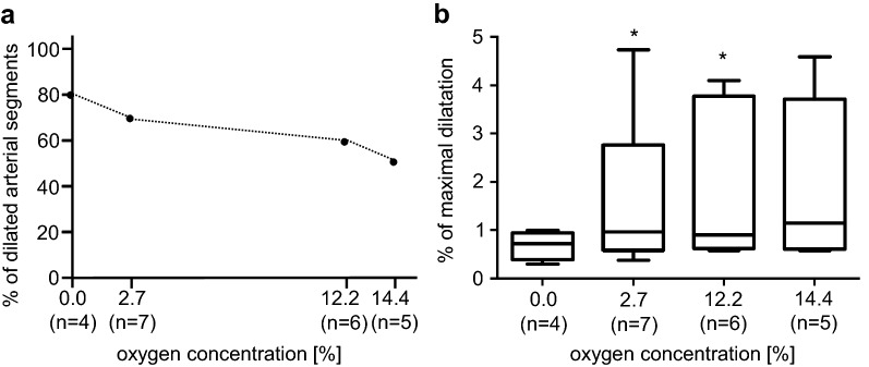
Results: Mild hypoxia 14.4% O2 induced, in 50% of mesenteric artery segments from wild-type (wt) mice, a vasodilatation; severe hypoxia recruited further segments responding with vasodilatation reaching 80% under anoxia. However, the extension of dilatation of luminal arterial diameter reduced from 1.96% ± 0.55 at 14.4% O2 to 0.68% ± 0.13 under anoxia. Arteries exposed to hypoxia in Ca2+-free solution responded to lower oxygen levels with increasing degree of vasodilatation (0.85% ± 0.19 at 14.4% O2 vs. 1.53% ± 0.42 at 2.7% O2). Inhibition of voltage-gated Ca2+-influx using NiCl2 completely diminished hypoxia-induced vasodilatation. Instead, all arterial segments investigated constricted. Furthermore, we did not observe altered hypoxia-induced vasomotion in eNOS-/-- or TASK1-/- mice compared to wt animals.
Conclusions: The present study demonstrated that hypoxic vasodilatation in mice mesenteric arteries is mediated by a NO-independent mechanism. In this experimental setting, we found evidence for Ca2+-mediated activation of ion channels causing hypoxic vasodilatation.
Keywords: Hypoxia; Mesenteric artery; Mice; Vasomotion.
Figures


Similar articles
-
Age-dependent impact of CaV 3.2 T-type calcium channel deletion on myogenic tone and flow-mediated vasodilatation in small arteries.J Physiol. 2016 Oct 15;594(20):5881-5898. doi: 10.1113/JP271470. Epub 2016 Feb 18. J Physiol. 2016. PMID: 26752249 Free PMC article.
-
Calcium-activated potassium channel and connexin expression in small mesenteric arteries from eNOS-deficient (eNOS-/-) and eNOS-expressing (eNOS+/+) mice.Eur J Pharmacol. 2007 Apr 10;560(2-3):193-200. doi: 10.1016/j.ejphar.2007.01.018. Epub 2007 Jan 19. Eur J Pharmacol. 2007. PMID: 17300779
-
T-type Ca(2+) channels facilitate NO-formation, vasodilatation and NO-mediated modulation of blood pressure.Pflugers Arch. 2014 Dec;466(12):2205-14. doi: 10.1007/s00424-014-1492-4. Epub 2014 Mar 14. Pflugers Arch. 2014. PMID: 24627154
-
Mechanisms of cellular synchronization in the vascular wall. Mechanisms of vasomotion.Dan Med Bull. 2010 Oct;57(10):B4191. Dan Med Bull. 2010. PMID: 21040688 Review.
-
Modulation of voltage-gated Ca2+ channels by O2 tension. Significance for arterial oxygen chemoreception.Adv Exp Med Biol. 1996;410:97-103. doi: 10.1007/978-1-4615-5891-0_14. Adv Exp Med Biol. 1996. PMID: 9030284 Review. No abstract available.
Cited by
-
Hif1α-dependent mitochondrial acute O2 sensing and signaling to myocyte Ca2+ channels mediate arterial hypoxic vasodilation.Nat Commun. 2024 Aug 5;15(1):6649. doi: 10.1038/s41467-024-51023-3. Nat Commun. 2024. PMID: 39103356 Free PMC article.
-
Hypoxic Conditions Promote Rhythmic Contractile Oscillations Mediated by Voltage-Gated Sodium Channels Activation in Human Arteries.Int J Mol Sci. 2021 Mar 4;22(5):2570. doi: 10.3390/ijms22052570. Int J Mol Sci. 2021. PMID: 33806419 Free PMC article.
-
Retinal capillary perfusion heterogeneity in diabetic retinopathy detected by optical coherence tomography angiography.Int J Retina Vitreous. 2024 Jan 25;10(1):12. doi: 10.1186/s40942-024-00528-6. Int J Retina Vitreous. 2024. PMID: 38273321 Free PMC article.
References
MeSH terms
Substances
LinkOut - more resources
Full Text Sources
Other Literature Sources
Miscellaneous
