. 2018;14(12):2139-2154.
doi: 10.1080/15548627.2018.1509607. Epub 2018 Sep 13.

Pathological missorting of endogenous MAPT/Tau in neurons caused by failure of protein degradation systems

Affiliations

Pathological missorting of endogenous MAPT/Tau in neurons caused by failure of protein degradation systems

Varun Balaji et al. Autophagy. 2018.

Abstract

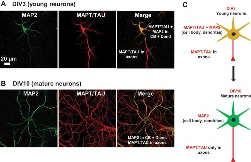
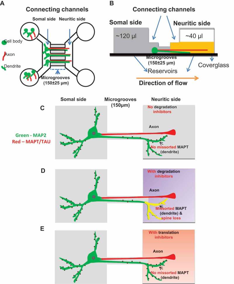
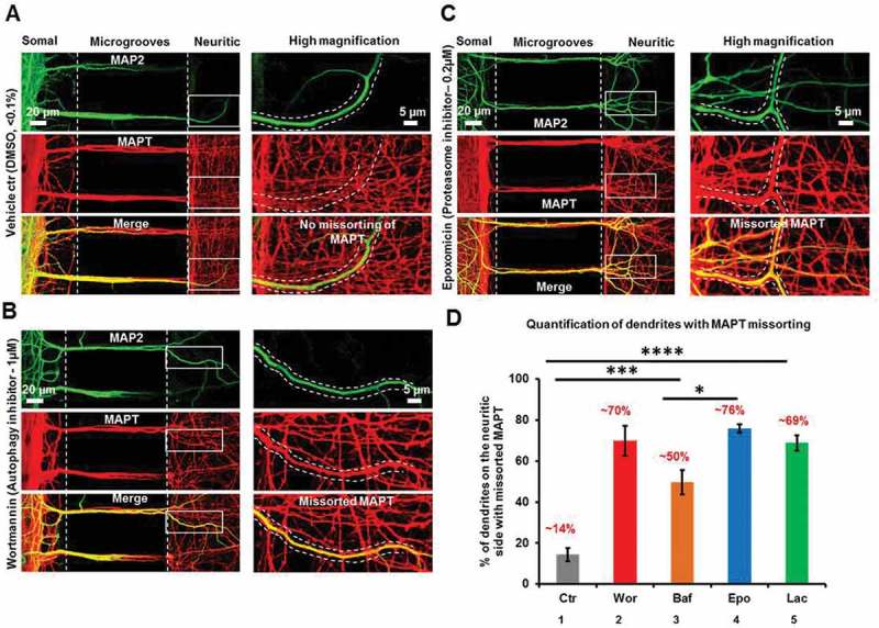
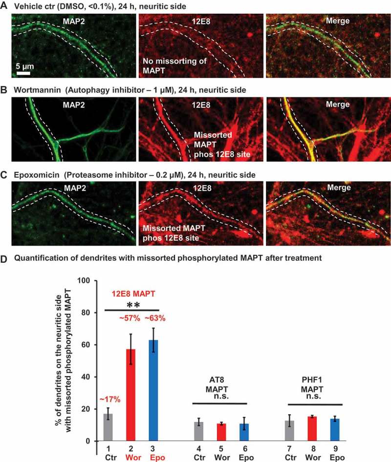
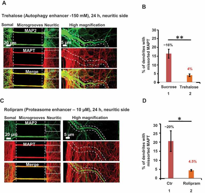
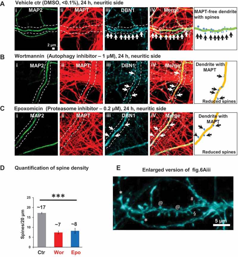
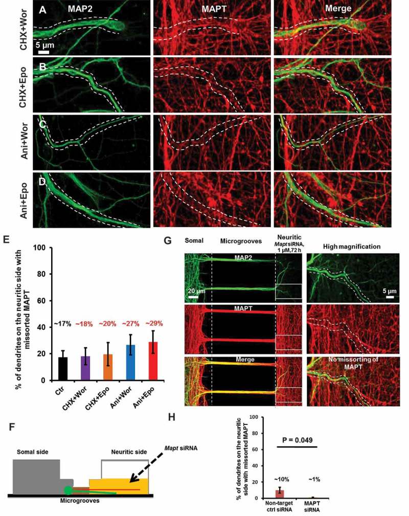
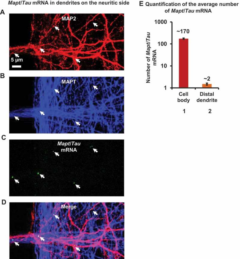
Missorting of MAPT/Tau represents one of the early signs of neurodegeneration in Alzheimer disease. The triggers for this are still a matter of debate. Here we investigated the sorting mechanisms of endogenous MAPT in mature primary neurons using microfluidic chambers (MFCs) where cell compartments can be observed separately. Blocking protein degradation pathways with proteasomal or autophagy inhibitors dramatically increased the missorting of MAPT in dendrites on the neuritic side, suggesting that degradation of MAPT in dendrites is a major determinant for the physiological axonal distribution of MAPT. Such missorted dendritic MAPT differed in its phosphorylation pattern from axonal MAPT. By contrast, enhancing autophagy or proteasomal pathways strongly reduced MAPT missorting, thereby confirming the role of protein degradation pathways in the polar distribution of MAPT. Dendritic missorting of MAPT by blocking protein degradation resulted in the loss of spines but not in overall cell toxicity. Inhibition of local protein synthesis in dendrites eliminated the missorting of MAPT, indicating that the accumulation of dendritic MAPT is locally generated. In support of this, a substantial fraction of Mapt/Tau mRNA was detected in dendrites. Taken together, our results indicate that the autophagy and proteasomal pathways play important roles in fine-tuning dendritic MAPT levels and thereby prevent synaptic toxicity caused by MAPT accumulation. Abbreviations Ani: anisomycin; Baf: bafilomycin A1; BSA: bovine serum albumin; cAMP: cyclic adenosine monophosphate; CHX: cycloheximide; DMSO: dimethyl sulfoxide; DIV: days in vitro; Epo: epoxomicin; E18: embryonic day 18; FISH: fluorescence in situ hybridization; IgG: immunoglobulin; kDa: kilodalton; Lac: lactacystin; LDH: lactate dehydrogenase; MFC: microfluidic chambers; MAPs: microtubule-associated proteins; MAPT/Tau: microtubule-associated protein tau; PVDF: polyvinylidene difluoride; PBS: phosphate-buffered saline; PRKA: protein kinase AMP-activated; RD150: round device 150; RT: room temperature; SDS: sodium dodecyl sulfate; SEM: standard error of the mean; Wor: wortmannin.

Keywords: Alzheimer disease; MAPT/Tau; autophagy; degradation; microfluidic chambers; proteasome.


Figures

Figure 1.
Figure 1.
Expression and localization of MAPT in developing neurons. (a) Distribution of MAPT in developing neurons determined by immunofluorescence. Rat hippocampal neurons cultured on coverglasses in 24-well plates were fixed and double stained for MAP2 (green) and MAPT (red). MAPT is distributed abundantly in the somata and processes of neurons at DIV3. (b) At DIV10, MAPT immunoreactivity disappears from the somatodendritic compartment and MAPT is sorted mainly to the axons. Scale bar: 20 µm. (c) Cartoon illustrating the transition in MAP2 and MAPT distribution from immature to mature neurons.
Figure 2.
Figure 2.
Separation of dendrites and axons from soma using microfluidic chambers (MFC). (a) Diagram of an MFC showing the somal (left) and neuritic sides (right) connected by microgrooves with a length of 150 ± 25 µm and a width of 10 µm. The somal side contains cell bodies, dendrites (green) and axons (red). The neuritic side contains only axons and dendrites. The microgrooves do not allow the entry of the cell bodies so that only the neurites can pass through from the somal to the neuritic side. (b) Schematic of the side view of an MFC showing fluidic isolation. The somal side is shaded in dark gray and the neuritic side in yellow. Fluidic isolation is achieved by removing a small volume of medium (~ 80 µl) from the neuritic side, generating a flow of liquid from the somal to the neuritic side (orange arrow). This prevents back diffusion of molecules from the neuritic to the somal side. (c-e) Schematics of the treatments and its effects in a neuron cultured in an MFC. In the absence of protein degradation inhibitors of autophagy and the proteasome, MAPT (red) sorts mainly into the axons and MAP2 (green) into the somatodendritic compartment (c). When protein degradation inhibitors are applied on the neuritic side, there is an increased fraction of dendrites on the neuritic side with MAPT accumulation (yellow) accompanied by loss of spines (d). In the presence of translation inhibitors on the neuritic side, no MAPT missorting into the dendrites is observed (e).
Figure 3.
Figure 3.
Inhibition of protein degradation (autophagy, proteasome) leads to MAPT missorting. Rat hippocampal neurons (DIV 21–25) cultured in microfluidic chambers were treated on the neuritic side for 24 h either with DMSO (control, a) or with the autophagy inhibitor (wortmannin [Wor], b) or the proteasomal inhibitor (epoxomicin [Epo], c). Dendrites were stained with MAP2 antibody (green) and total MAPT with K9JA antibody (red). Magnified images of the insets are shown on the right with a pair of eye-guiding dotted lines highlighting a dendrite with or without MAPT. (a) In the vehicle-treated control (DMSO, < 0.1%), MAPT is predominantly localized to the axons (see merged images at the bottom in a). Only a small fraction of dendrites colocalizes with MAPT. (b and c) In cultures treated with wortmannin (b, 1 µM, 24 h) or with epoxomicin (c, 0.2 µM, 24 h), the fraction of dendrites with MAPT increases strongly (see quantification in D) where a clear colocalization of MAPT with MAP2 (merged images at the bottom in b and c) becomes visible. Scale bars in all overview images on left: 20 µm; in all magnifications of insets on right: 5 µm. (d) Quantification of dendrites on the neuritic side showing colocalization of MAPT with MAP2 following treatment with DMSO (ctr, bar 1) or with the autophagy (bars 2 and 3) or proteasomal (bars 4 and 5) inhibitors. (n > 180 dendrites/dendritic branches from 3–6 experiments; one-way ANOVA with Tukey’s post hoc test; F [4,17] = 30.09; *p < 0.05; ***p < 0.001; ****p < 0.0001).
Figure 4.
Figure 4.
Missorting of MAPT into dendrites coincides with elevated phosphorylation at the 12E8 epitope, but not at the AT8 or PHF1 sites. Rat hippocampal neurons (DIV 21–25) cultured in microfluidic chambers treated on the neuritic side for 24 h either with DMSO (control, a), or with the autophagy inhibitor wortmannin (b) or proteasomal inhibitor epoxomicin (c). Phosphorylation-dependent MAPT antibody 12E8 was used to probe the phosphorylation state of MAPT at S262/S356 residues and MAP2 antibody was used to highlight dendrites. Images of the dendrites on the neuritic side are shown highlighting a dendrite with or without phospho-MAPT. (a-c) In vehicle-treated control (DMSO, < 0.1%) MAPT sorts mainly to the axons (a). Treatment with wortmannin (1 µM, 24 h) or epoxomicin (0.2 µM, 24 h) on the neuritic side causes an increase of MAPT, in the dendrites, which is phosphorylated at the 12E8 site (b and c). Scale bar: 5 µm. (d) Quantification of dendrites on the neuritic side showing colocalization of MAP2 with the phospho-MAPT antibodies 12E8 (n > 700 dendrites/dendritic branches from 5 experiments; one-way ANOVA with Tukey’s post hoc test; F [2,12] = 12; **p < 0.01), AT8 (n = > 70 dendrites/dendritic branches from 3 experiments; one-way ANOVA with Tukey’s post hoc test; F [2,6] = 0.05; p = 0.09) or PHF1 (n = > 30 dendrites/dendritic branches 3 experiments; one-way ANOVA with Tukey’s post hoc test; F [2,6] = 0.31; p = 0.74). ns, not significant.
Figure 5.
Figure 5.
Enhancement of autophagy or proteasome activity reduces MAPT missorting. Rat hippocampal neurons (DIV 21–25) cultured in MFCs were treated on the neuritic side with sucrose (as control) or with trehalose, an enhancer of autophagy (a) or with rolipram, an enhancer of the proteasome (c) for 24 h. The dendrites are stained with MAP2 antibody (green) and MAPT is stained with K9JA antibody (red). Magnified images of the insets are shown on the right with a pair of eye-guiding dotted lines highlighting a dendrite without MAPT. (a and b) Impact of stimulation of autophagy on missorting of MAPT. In cultures treated with trehalose (150 mM, 24h) on the neuritic side, the fraction of dendrites with MAPT decreases strongly (see quantification in B) and a more stringent localization to axons is seen (a). Treatment with sucrose (150 mM, 24 h) resulted in a similar fraction of dendrites with missorted MAPT as DMSO treated control cultures, but trehalose (150 mM, 24 h) strongly reduced missorting of MAPT 4 fold (~ 4%) (b). In A, the image of the dendrites on the neuritic side represent reduced colocalization of MAPT with MAP2 (see panels representing high magnification of insets). Scale bars in all overview images on left:20 µm; in all magnifications of insets on right: 5 µm. Error bars, SEM from n > 450 dendrites/dendritic branches from 4 experiments. **< 0.005, using Student’s t test. (c and d) Impact of stimulation of proteasome on missorting of MAPT. In cultures treated with rolipram (10 µM, 24 h) on the neuritic side, the fraction of dendrites with MAPT decreases by ~ 70% (see quantification in d). In c, the image of the dendrites on the neuritic side represents reduced colocalization of MAPT with MAP2 (see high magnification of insets). Error bars, SEM from n > 280 dendrites/dendritic branches from 4 experiments. *< 0.05, using Student’s t test.
Figure 6.
Figure 6.
Local treatment with protein degradation inhibitors leads to dendritic MAPT mislocalization and spine loss. Rat hippocampal neurons (DIV 21–25) cultured in microfluidic chambers were treated on the neuritic side for 24 h either with DMSO (control, a), with the autophagy inhibitor wortmannin (b) or with the proteasomal inhibitor epoxomicin (c). MAP2 antibody (green), K9JA antibody (red) and DBN1 (cyan) were used to label dendrites, MAPT and spines, respectively. Only magnified images of the neuritic side are shown here. (a) In control, neurons treated with vehicle at the neuritic side (DMSO, < 0.1%, 24 h), MAPT is predominantly localized to the axons and the dendrites have a normal spine distribution and morphology (Aiii) (arrows). (b and c) In cultures treated with wortmannin (b, 1 µM, 24 h) or with epoxomicin (c, 0.2 µM, 24 h) MAPT can be observed in dendrites, and these dendrites show reduction of spines, and the remaining spines become stubby in nature (Biii and Ciii). Scale bar: 2 µm. (d) Quantification of the spine density of the dendrites on the neuritic side after treatment with DMSO or with protein degradation inhibitors (n = 17–20 dendrites from 3 experiments; one-way ANOVA with Tukey’s post hoc test; F [2,6] = 39.29; ***p < 0.001). (e) Enlarged version of Figure 6(a)iii clearly demonstrates different types of spine morphology such as mushroom head (*), stubby(@), thin(#) and filopodia(§) spines.
Figure 7.
Figure 7.
Protein translation inhibitors prevent MAPT missorting. Rat hippocampal neurons (DIV 21–25) cultured in MFCs treated on the neuritic side with the protein translation inhibitors cycloheximide (CHX) or anisomycin (Ani) combined either with wortmannin (a and c) or with epoxomicin (b and d) for 24 h or with Mapt siRNA (F-H) for 72 h. The dendrites are stained with MAP2 antibody (green) and MAPT is stained with K9JA antibody (red). Images of the dendrites on the neuritic side are shown highlighting a dendrite without MAPT. (a and b) When the neurons were treated with cycloheximide (10 µM) together with the autophagy inhibitor wortmannin (1 µM) (a) or with the proteasomal inhibitor epoxomicin (0.2 µM) (b) for 24 h, there was no increase in the dendritic accumulation of MAPT (quantification in e). (c and d) Similar experiment to A, B using anisomycin (10 µM) combined with wortmannin (1 µM) (c) or epoxomicin (0.2 µM) (d). There was no increase in the dendritic accumulation of MAPT (quantification in E). Scale bar: 5 µm. (e) Quantification of dendrites on the neuritic side showing colocalization of MAP2 with MAPT (n > 180 dendrites/dendritic branches from 3–5 experiments; one-way ANOVA with Tukey’s post hoc test; F [4,15] = 0.58; p = 0.68). (f–h) When the neurons were treated with siRNA against Mapt in the neuritic side (1 µM, 72 h) (f) there was very low accumulation of MAPT in dendrites on the neuritic side (see merged images in the right panels [g] and quantification [h]). Scale bars in all overview images on left: 20 µm; in all magnifications of insets on right: 5 µm. Error bar, SEM from n = > 800 dendrites/dendritic branches from 4 experiments.
Figure 8.
Figure 8.
Fluorescence in situ hybridization reveals the majority of Mapt/Tau mRNA in the cell body with a minority in distal dendrites. (a-d) Mapt/Tau mRNA in rat hippocampal neurons (DIV 21–25) cultured in MFCs monitored by FISH with a rat Mapt/Tau mRNA probe. Axons and dendrites were visualized by immunostaining with pan-MAPT antibody K9JA (blue, b) and anti-MAP2 antibody (red, a) respectively. Images of the dendrites on the neuritic side are shown. Note that Mapt/Tau mRNA (green puncta, c) can be found in distal dendrites. Arrows indicate a sparse distribution of Mapt/Tau mRNA in distal dendrites. Scale bar: 5 µm. (e) Quantification of the average number of Mapt/Tau mRNA puncta in the cell body (~ 170) vs a distal dendrite (~ 2). Error bars, SEM from n = 45 cell bodies and 106 distal dendrites from 3 individual experiments.

References

    1. Mandelkow EM, Mandelkow E.. Biochemistry and cell biology of tau protein in neurofibrillary degeneration. Cold Spring Harb Perspect Med. 2012. July;2(7):a006247 PubMed PMID: 22762014; PubMed Central PMCID: PMC3385935. - PMC - PubMed
    1. Drubin DG, Kirschner MW. Tau protein function in living cells. J Cell Biol. 1986. December;103(6 Pt 2):2739–2746. PubMed PMID: 3098742; PubMed Central PMCID: PMC2114585. - PMC - PubMed
    1. Binder LI, Frankfurter A, Rebhun LI. The distribution of tau in the mammalian central nervous system. J Cell Biol. 1985. October;101(4):1371–1378. PubMed PMID: 3930508; PubMed Central PMCID: PMC2113928. - PMC - PubMed
    1. Kosik KS, Joachim CL, Selkoe DJ. Microtubule-associated protein tau (tau) is a major antigenic component of paired helical filaments in Alzheimer disease. Proc Natl Acad Sci USA. 1986. June;83(11):4044–4048. PubMed PMID: 2424016; PubMed Central PMCID: PMC323662. - PMC - PubMed
    1. Braak E, Braak H, Mandelkow EM. A sequence of cytoskeleton changes related to the formation of neurofibrillary tangles and neuropil threads. Acta Neuropathol. 1994;87(6):554–567. PubMed PMID: 7522386. - PubMed

Publication types

MeSH terms