. 2018 Sep 6;174(6):1477-1491.e19.
doi: 10.1016/j.cell.2018.07.041. Epub 2018 Aug 23.

TBK1 Suppresses RIPK1-Driven Apoptosis and Inflammation during Development and in Aging

Affiliations

TBK1 Suppresses RIPK1-Driven Apoptosis and Inflammation during Development and in Aging

Daichao Xu et al. Cell. .

Abstract

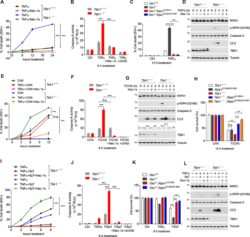
Aging is a major risk factor for both genetic and sporadic neurodegenerative disorders. However, it is unclear how aging interacts with genetic predispositions to promote neurodegeneration. Here, we investigate how partial loss of function of TBK1, a major genetic cause for amyotrophic lateral sclerosis (ALS) and frontotemporal dementia (FTD) comorbidity, leads to age-dependent neurodegeneration. We show that TBK1 is an endogenous inhibitor of RIPK1 and the embryonic lethality of Tbk1-/- mice is dependent on RIPK1 kinase activity. In aging human brains, another endogenous RIPK1 inhibitor, TAK1, exhibits a marked decrease in expression. We show that in Tbk1+/- mice, the reduced myeloid TAK1 expression promotes all the key hallmarks of ALS/FTD, including neuroinflammation, TDP-43 aggregation, axonal degeneration, neuronal loss, and behavior deficits, which are blocked upon inhibition of RIPK1. Thus, aging facilitates RIPK1 activation by reducing TAK1 expression, which cooperates with genetic risk factors to promote the onset of ALS/FTD.

Keywords: ALS; FTD; RIPK1; RIPK1-dependent apoptosis; TAK1; TBK1; caspase; necroptosis.


Conflict of interest statement

Declaration of Interests

JY is a consultant of Denali Therapeutics.

Figures

Figure 1.
Figure 1.. Inhibition of RIPK1 kinase activity blocks embryonic lethality of Tbk1−/− mice.
(A) Numbers of offspring from intercrossing Tbk1+/−;Ripk1D138N/D138N parents. (B) Histological analysis and TUNEL assays were performed on E13.5 liver sections (n=3). H&E, haematoxylin and eosin. (C and D) The liver samples (E13.5) were analyzed by immunoblotting (C) and immunostained for cleaved caspase-3 (CC3), RIPK1 or DAPI for nuclei (n=3) (D). (E) Quantitative RT-PCR analysis of the mRNA expression of cytokines and chemokines in E13.5 pups’ liver (n=9, mean ± SEM). ( F) The numbers and genotypes of weaned offspring from Tbk1+/−;Ripk3−/− parents. (G) p-RIPK3 immunohistostaining (brown, n=3). Microscopic quantification of p-RIPK3 positive cells on liver sections (E13.5) (right). (mean ± SEM. **p < 0.01, ***p < 0.001). See also Figures S1 and S2.
Figure 2.
Figure 2.. TBK1 deficiency promotes RIPK1-dependent apoptosis.
(A-D) MEFs were treated with 10 ng/ml mTNFα, +/or - Nec-1s (10 μM). Cell death (A, C) and caspase-8 activity (B) were measured by SytoxGreen positivity and Caspase-Glo 8 assay, respectively. The levels of p-RIPK1(S166) and CC3 were determined by immunoblotting (D). Data are represented as mean ± SD. (E-H) MEFs of indicated genotypes were treated with 1 μM CHX for 0.5 h followed by mTNFα, +/or - Nec-1s. Cell death was measured by SytoxGreen positivity (E) or CellTiter-Glo assay (H). Caspase-8 activity was measured by Caspase-Glo 8 assay (F). The levels of p-RIPK1(S166) and CC3 were determined by immunoblotting (G). Data are represented as mean ± SD. (I-L) MEFs were treated with 100 nM (5Z)-7-Oxozeaeno for 0.5 h followed by mTNFα, +/or - Nec-1s. Cell death was measured by SytoxGreen positivity (I) or CellTiter-Glo assay (K). Caspase-8 activity was measured by Caspase-Glo 8 assay (J). The levels of p-RIPK1(S166) and CC3 were determined by immunoblotting (L). Data are represented as mean ± SD. ***p < 0.001, n.s. not significant. See also Figure S3.
Figure 3.
Figure 3.. TBK1 is recruited into TNF-RSC to suppress RIPK1 activation.
(A-C) MEFs were stimulated by Flag-mTNFα (100 ng/ml), and SM164 (50 nM) or Nec-1s. TNF-RSC (Complex I) was immunoprecipitated using anti-Flag resin. The recruitment of TBK1 and RIPK1 were analyzed by immunoblotting. TNFR1 was a control for TNF-RSC. (D-F) MEFs were stimulated by Flag-mTNFα with or without Nec-1s. TNF-RSC was immunoprecipitated using anti-Flag resin. The lysates were analyzed by immunoblotting using anti-p-RIPK1(S166) antibody. (G) WT and Tbk1−/− MEFs were pre-incubated with 20 μM zVAD-fmk, +/or - Nec-1s for 0.5 h and then stimulated with mTNFα. The complex II was isolated by FADD immunoprecipitated and RIPK1 binding was revealed by immunoblotting. See also Figure S4.
Figure 4.
Figure 4.. TBK1 inhibits RIPK1 by direct phosphorylation.
(A) The structural comparison of the RIPK1 kinase domain (PDB ID: 4ITI) and the PKC/Par3 complex (PDB ID: 4ITI). The kinase domain of PKC and the bound Par3 peptide in the PKC/Par3 complex are shown in grey and in orange respectively, while the RIPK1 kinase domain is drawn in blue; the side chain of S1060 phosphorylation site of Par3 is highlighted and displayed in the stick-ball model, and the side chains of the related key residues are shown in the stick mode; while the relevant hydrogen bonds are indicated by black dash lines. (B) A schematic diagram of inducible heterodimerization of mouse RIPK1 DD-DmrC-HA-GFP fusion with RIPK1 DD-DmrA-Myc fusion. (C and D) HEK293T cells were co-transfected with expression vectors for mRIPK1 DD(WT)-DmrC-HA-GFP, mRIPK1 DD(WT)-DmrA-Myc or mRIPK1 DD(D138N)-DmrA-Myc (C), mRIPK1 DD(WT)-DmrC-HA-GFP, mRIPK1 DD(WT)-DmrA-Myc, mRIPK1 DD(T190E)-DmrA-Myc or mRIPK1 DD(T190A)-DmrA-Myc (D) as indicated for 12h, and then treated with 100nM A/C Heterodimerizer ligand (AP21967) for different periods of time as indicated. The levels of p-RIPK1 (S166) were analyzed by immunoblotting. (E) Jurkat cells were pre-treated with or without MRT67307 for 0.5 h and then stimulated with 10 ng/ml hTNFα. RIPK1 was then immunoprecipitated with p-RIPK1(T189) ab and determined by immunoblotting. (F) HEK293T cells were transfected with WT, T190E, T190A or K45M mutant Flag-hRIPK1 and treated with Nec-1s for 20 h. Flag-RIPK1 was then immunoprecipitated using anti-Flag and incubated with or without 100 μM ATP or 50 μM Nec-1s as indicated at 30°C for 30min. The samples were analyzed by immunoblotting with p-RIPK1(S166). (G) Tbk1−/−;Ripk1CrisprKO MEFs were retrovirally reconstituted with HA tagged WT, T190E or T190A mutant RIPK1. Reconstituted cells were stimulated with TNFα. Cell death was measured by ToxiLight (n=4. Mean ± SD. **p < 0.01, ***p < 0.001). See also Figure S5.
Figure 5.
Figure 5.. Over-activation of TAK1 in TBK1 deficient cells and reduction of TAK1 in human aging brains.
(A) WT and Tbk1−/− MEFs were stimulated with mTNFα and analyzed by immunoblotting. (B) Tbk1+/+ and Tbk1−/− MEFs were treated with TNFα for 15 min and then were immunoprecipitated with TAK1 ab. The precipitated TAK1 (IP: anti-TAK1) kinase activity was measured by incubating the kinase with 4 μg GST fused mouse TAK1 peptide (aa180–195) in vitro in the presence of ATP or 5z7 at 30 °C for 30 min. The samples were analyzed by immunoblotting with p-TAK1(T184/T187). (C) Immunoblotting analysis of human frontal cortex (12 young and 12 older individuals) (top) and the quantification of TAK1 levels (bottom) (mean ± SEM). (D) MEFs of indicated genotypes were stimulated by Flag-mTNFα. TNF-RSC was immunoprecipitated using anti-FLAG resin. The activation of RIPK1 was analyzed by immunoblotting using anti-p-RIPK1(S166) ab. (E-G) MEFs were treated with mTNFα, +/or - Nec-1s. Cell death was measured by CellTiter-Glo assay (E, F) (mean ± SD). The levels of CC3 were determined by immunoblotting (G). *p < 0.05, **p < 0.01, ***p < 0.001. See also Figure S6.
Figure 6.
Figure 6.. Double heterozygosity of TBK1 and TAK1 promotes microglia activation.
(A) Immunostaining of microglia marker IBA1 in the ventral spinal cords and cortex from 6 months old mice and quantification (50 slices/genotype) (mean ± SD). (B) MHC-II cytometry analysis of microglia (CD11b+LY6CCD45IntermediateCX3CR1+) in cerebrum and spinal cord of WT, Tbk1+/−;Tak1 ΔM/+ and Tbk1+/−;Tak1 ΔM/+;Ripk1D138N/+ mice (male, 6 months old; n = 2 per group). (C) GO analysis of genes that are either up-regulated in Tbk1+/−;Tak1 ΔM/+ microglia (D) or down- regulated in Tbk1+/−;Tak1 ΔM/+ microglia in RIPK1-dependent fashion (E). (F) The cytokine profiles in the spinal cords from 6 months old mice were measured by quantitative PCR (n=3, mean ± SEM). *p < 0.05, **p < 0.01, ***p < 0.001. See also Figure S6.
Figure 7.
Figure 7.. TBK1/TAK1 double heterozygosity promotes axonal defects in spinal cords and FTD phenotype in the CNS.
(A) EM analysis of motor axonal myelination in the ventrolateral lumbar spinal cords from 6 months old mice. (B-D) The mean axonal numbers, mean g-ratios, and mean axonal diameters (B); individual axonal diameter distribution (C); and g-ratio distribution (D) in the ventrolateral lumbar spinal cord white matter of mice (mean ± SEM). (E) The number of TUNEL+ cells in the lumbar spinal cords of indicated genotype (15 sections from 3 mice for each genotype) (mean ± SEM). (F and G) Mice (6 months old. WT, n=10; Tbk1+/−, n=11; Tak1 ΔM/+, n=9; Tbk1+/−;Tak1 ΔM/+, n=10; Tbk1+/−;Tak1 ΔM/+;Ripk1D138N/+, n=9) were tested in open-field. The total distance traveled showed no difference (F). Tbk1+/−;Tak1 ΔM/+ mice, but not Tbk1+/− or Tak1 ΔM/+ mice, showed a significant deficit on the vertical rearing activity, which was blocked in Tbk1+/−;Tak1 ΔM/+;Ripk1D138N/+ mice (G) (mean ± SD). (H) Representative images of NeuN-labeled cells in the cortex of WT mice (n=3), Tbk1+/−;Tak1 ΔM/+ mice (n=3) and Tbk1+/−;Tak1 ΔM/+;Ripk1D138N/+ mice (n=3) and quantification (right) (mean ± SEM). (I) The average percentage of cells with TDP-43 inclusions in mice of indicated genotypes was imaged and quantified (500 cells per mouse, n=3 mice). (J and K) Anxiety-related behavior was determined by bright open field test by quantifing the distance travelled in the center area normalized to total distance travelled (J), and elevated plus maze showing the percent of time spent on the open arm (K). (WT, n=10; Tbk1+/−, n=11; Tak1 ΔM/+, n=9; Tbk1+/−;Tak1 ΔM/+, n=10; Tbk1+/−;Tak1 ΔM/+;Ripk1D138N/+, n=10) (mean ± SD. *p < 0.05, **p < 0.01, ***p < 0.001, n.s. not significant). See also Figure S7.

Comment in

References

    1. Barron KD (1995). The microglial cell. A historical review. Journal of the neurological sciences 134 Suppl, 57–68. - PubMed
    1. Bayle JH, Grimley JS, Stankunas K, Gestwicki JE, Wandless TJ, and Crabtree GR (2006). Rapamycin analogs with differential binding specificity permit orthogonal control of protein activity. Chemistry & biology 13, 99–107. - PubMed
    1. Berger SB, Kasparcova V, Hoffman S, Swift B, Dare L, Schaeffer M, Capriotti C, Cook M, Finger J, Hughes-Earle A, et al. (2014). Cutting Edge: RIP1 kinase activity is dispensable for normal development but is a key regulator of inflammation in SHARPIN-deficient mice. J Immunol 192, 5476–5480. - PMC - PubMed
    1. Bonnard M, Mirtsos C, Suzuki S, Graham K, Huang J, Ng M, Itie A, Wakeham A, Shahinian A, Henzel WJ, et al. (2000). Deficiency of T2K leads to apoptotic liver degeneration and impaired NF-kappaB-dependent gene transcription. EMBO J 19, 4976–4985. - PMC - PubMed
    1. Caccamo A, Branca C, Piras IS, Ferreira E, Huentelman MJ, Liang WS, Readhead B, Dudley JT, Spangenberg EE, Green KN, et al. (2017). Necroptosis activation in Alzheimer’s disease. Nat Neurosci 20, 1236–1246. - PubMed

Publication types

MeSH terms

Substances