. 2018 Sep 12;24(3):439-446.e4.
doi: 10.1016/j.chom.2018.08.001. Epub 2018 Aug 23.

Alveolar Macrophages Provide an Early Mycobacterium tuberculosis Niche and Initiate Dissemination

Affiliations

Alveolar Macrophages Provide an Early Mycobacterium tuberculosis Niche and Initiate Dissemination

Sara B Cohen et al. Cell Host Microbe. .

Abstract

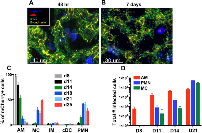
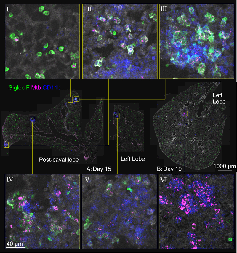
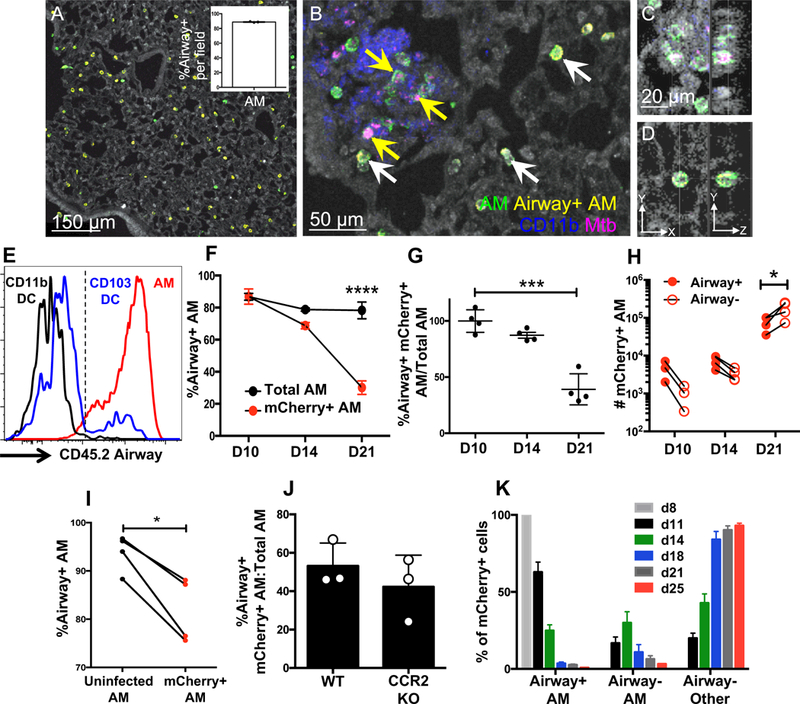
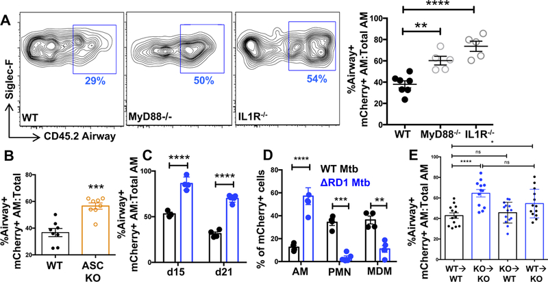
Mycobacterium tuberculosis (Mtb) infection is initiated in the distal airways, but the bacteria ultimately disseminate to the lung interstitium. Although various cell types, including alveolar macrophages (AM), neutrophils, and permissive monocytes, are known to be infected with Mtb, the initially infected cells as well as those that mediate dissemination from the alveoli to the lung interstitium are unknown. In this study, using a murine infection model, we reveal that early, productive Mtb infection occurs almost exclusively within airway-resident AM. Thereafter Mtb-infected, but not uninfected, AM localize to the lung interstitium through mechanisms requiring an intact Mtb ESX-1 secretion system. Relocalization of infected AM precedes Mtb uptake by recruited monocyte-derived macrophages and neutrophils. This dissemination process is driven by non-hematopoietic host MyD88/interleukin-1 receptor inflammasome signaling. Thus, interleukin-1-mediated crosstalk between Mtb-infected AM and non-hematopoietic cells promotes pulmonary Mtb infection by enabling infected cells to disseminate from the alveoli to the lung interstitium.

Keywords: ESX-1; IL-1; alveolar macrophages; granuloma; innate immunity; lung; pulmonary tuberculosis.


Conflict of interest statement

Declaration of interests. The authors have no conflicts of interest to declare.

Figures

Figure 1
Figure 1. See also Figure S1. AM are the major target of Mtb early post-infection.
(A,B) Imaging of infected alveolar macrophages (AM, SiglecF+) from lung sections collected at (A) 48 h (7 sections, n=5 mice) or (B) D7–10 (4 sections, n=3 mice). (C) Composition of mCherry+ lung leukocytes over a timecourse of infection (3 pooled experiments, n=3–11 mice/timepoint). (D) Total numbers of infected AM, neutrophils (PMN), and monocyte-derived cells (MC) in the lung. Shown is a representative experiment (n=3–4 mice/timepoint) performed 3 times. The data are presented as means ± SEM.
Figure 2
Figure 2. See also Figure S2. Mtb-infected AM are found in the lung interstitium in the context of early cellular aggregates.
Representative immunofluorescence for Siglec F and CD11b at D15 (panels I-V) and D19 (panel VI) p.i. Zoom-in panels demonstrate infectious foci in various stages of progression: infected AM in alveolar sacs (panel I); co-localization of infected AMs with MC, with infection predominantly in AM (panel II); clustering of AM, with increased infection of MC and apparent interstitial localization of these cells (panels III, IV); progression of infection to MC, with AM pushed to the periphery (panel V). At D19, nearly all infected cells are MC (panel VI). Lung architecture is shown by autofluorescence (gray).
Figure 3
Figure 3. See also Figure S3. Intratracheal antibody administration enables tracking and quantification of airway-resident AM upon Mtb infection.
(A) Representative CD45.2 airway labeling of D15 p.i. B6 mice in an Mtb-free region with healthy lung architecture (autofluorescence). The inset shows the percentage of CD45.2+ AM over 3 independent fields. (B) A representative D15 Mtb-infected lung region, showing infected AM that are airway- and interstitial (yellow arrows) alongside airway+ uninfected AM in an alveolar pocket (white arrows). (C,D) Z-stack of D15 airway- (C) and airway+ (D) AM. (E) Airway labeling of AM, CD11b+ DC, and CD103+ DC in naive B6 mice by FACS. (F) Airway labeling of mCherry+ and total AM at indicated timepoints (n=4 mice/timepoint). (G) The data are plotted as ratios of mCherry+ over total AM to normalize for labeling efficiency. (H) Total numbers of airway+ and airway- infected AM over time (n=3 mice/timepoint). (I) Total airway+ AM were sorted from the lungs of D13 p.i. mice (n=10 mice) and adoptively transferred intratracheally into infection-matched congenic (B6.SJL) recipients (n=1 or 2 mice/group). Shown is the airway labeling of AM 3–5 days post-transfer from 3 independent experiments (each dot represents one mouse receiving AM pooled from 10 mice). (J) Airway labeling of infected AM in B6 and CCR2−/− mice D15 p.i. (n=3 mice/group). (K) Distribution of airway+ AM, airway- AM, and airway- other leukocytes among lung mCherry+ cells over time (3 pooled experiments, n=3–11 mice/timepoint). Multiple group comparisons were performed by 2-way ANOVA (F and H) and single group comparisons by unpaired t-test (G and J) or paired t-test (I). The data are presented as means ± SEM. All experiments were performed at least 2–3 times.
Figure 4
Figure 4. See also Figure S4. Lung interstitial localization of Mtb-infected AM requires inflammasome signaling on non-hematopoietic cells.
(A) Representative FACS plots and cumulative data of airway+ mCherry+ AM in B6, MyD88−/−, and IL1R−/− mice D19 p.i. (n=5 mice/group). (B) Cumulative data of airway labeling of B6 and ASC−/− mCherry+ AM D19 p.i. (n=8 mice/group from 2 pooled experiments). (C) Cumulative airway labeling of AM infected with either WT (H37Rv) or RD1-deficient Mtb (ΔRD1) at D15 and D21 p.i. (D) D21 composition of lung mCherry+ cells following H37Rv or ΔRD1 infection (n=4–5 mice/group). (E) Cumulative airway labeling data of infected AM in criss-cross IL1R−/− chimeras d19 p.i. (3 pooled experiments with 13–14 mice/group). Multiple-group comparisons were performed by 2-way ANOVA (D and E) and single group comparisons by t-test (A, B, and E). Experiments were performed at least twice and are shown either as representative data (A, C, and D) or pooled data from multiple experiments (B and E). The data are presented as means ± SEM.

Comment in

References

    1. Adams KN, Takaki K, Connolly LE, Wiedenhoft H, Winglee K, Humbert O, Edelstein PH, Cosma CL, and Ramakrishnan L (2011). Drug tolerance in replicating mycobacteria mediated by a macrophage-induced efflux mechanism. Cell 145, 39–53. - PMC - PubMed
    1. Barry KC, Fontana MF, Portman JL, Dugan AS, and Vance RE (2013). IL-1 alpha signaling initiates the inflammatory response to virulent Legionella pneumophila in vivo. J Immunol 190, 6329–6339. - PMC - PubMed
    1. Bermudez LE, Sangari FJ, Kolonoski P, Petrofsky M, and Goodman J (2002). The efficiency of the translocation of Mycobacterium tuberculosis across a bilayer of epithelial and endothelial cells as a model of the alveolar wall is a consequence of transport within mononuclear phagocytes and invasion of alveolar epithelial cells. Infect Immun 70, 140–146. - PMC - PubMed
    1. Cambier CJ, O’Leary SM, O’Sullivan MP, Keane J, and Ramakrishnan L (2017). Phenolic Glycolipid Facilitates Mycobacterial Escape from Microbicidal Tissue-Resident Macrophages. Immunity 47, 552–565 e554. - PMC - PubMed
    1. Cambier CJ, Takaki KK, Larson RP, Hernandez RE, Tobin DM, Urdahl KB, Cosma CL, and Ramakrishnan L (2014). Mycobacteria manipulate macrophage recruitment through coordinated use of membrane lipids. Nature 505, 218–222. - PMC - PubMed

Publication types

MeSH terms

Substances