. 2018 Sep 24;46(6):706-719.e5.
doi: 10.1016/j.devcel.2018.08.001. Epub 2018 Aug 23.

Spatially Restricted Regulation of Spätzle/Toll Signaling during Cell Competition

Affiliations

Spatially Restricted Regulation of Spätzle/Toll Signaling during Cell Competition

Lale Alpar et al. Dev Cell. .

Abstract

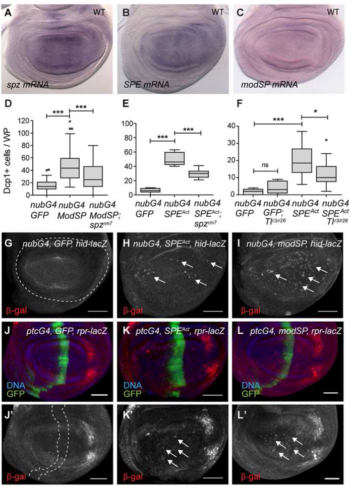
Cell competition employs comparisons of fitness to selectively eliminate cells sensed as less healthy. In Drosophila, apoptotic elimination of the weaker "loser" cells from growing wing discs is induced by a signaling module consisting of the Toll ligand Spätzle (Spz), several Toll-related receptors, and NF-κB factors. How this module is activated and restricted to competing disc cells is unknown. Here, we use Myc-induced cell competition to demonstrate that loser cell elimination requires local wing disc synthesis of Spz. We identify Spz processing enzyme (SPE) and modular serine protease (ModSP) as activators of Spz-regulated competitive signaling and show that "winner" cells trigger elimination of nearby WT cells by boosting SPE production. Moreover, Spz requires both Toll and Toll-8 to induce apoptosis of wing disc cells. Thus, during cell competition, Spz-mediated signaling is strictly confined to the imaginal disc, allowing errors in tissue fitness to be corrected without compromising organismal physiology.

Keywords: Drosophila; Myc; Spätzle; Toll receptors; cancer; cell competition; growth and development; inflammation; wing imaginal disc; “cheaters”.


Conflict of interest statement

Declaration of Interests

The authors declare no competing interests.

Figures

Figure 1:
Figure 1:. Spz and Toll are required for cell competition in wing imaginal discs.
(A) Schematic of clonal assay for Myc-induced cell competition. Clones generated by ‘Flp-out’ of an FRT (>) flanked cassette in tub>CD2>Gal4 (left) or tub>myc>Gal4 (right), marked by Gal4 controlled green fluorescent protein (GFP) expression. Neutral, GFP+ clones generated in a WT background (left) are controls for clone growth in a non-competitive context. GFP+ clones of WT cells surrounded by tub>myc>Gal4 cells become ‘losers’. (B-D) Wing discs with non-competing control clones marked by nuclear (n) GFP (B), UAS-nGFP+ loser clones (C), and UAS-CD8-GFP+ loser clones in the null spzKG05402 background (D). (E-F) Results of competition assays in (E) Tl and (F) spz null larvae. Tukey plot of (median and quartile) size of control clones in WT and mutant larvae (white), loser clones in WT larvae (light grey,) and loser clones in Tl or spz null larvae (dark grey). Clone number scored/genotype are in box plots. *** P<0.0005, ** P<0.005, * P<0.05 (unpaired t-test with Welch’s correction). (G) Results of qPCR showing spz expression in wing discs (WD), central nervous system (CNS), fat body (FB), salivary glands (SG), gut (G) and hemocytes (HC) of 3rd instar larvae. Error bars, SD. (See also Fig. S1).
Figure 2:
Figure 2:. Local expression of spz is required to eliminate loser cells in wing discs.
(A-B) Anti-mCherry staining of Spz-mCherry expression in wing discs from (A) 96 hr or (B) 72hr larvae. A and A’ show apical and medial sections, respectively, from the same disc. F-actin labeling marks the apical cell surface. (A” and B’) Z-sections of the wing discs (positions shown by red dashed lines on xy-plane images). Images are sum projections of multiple sections. Scale bars, 50 m. (C) Schematic of tub>myc>lexA/lexO competition assay with tissue-specific spz-RNAi knock-down. (D-E) Tukey box plots show clone size distribution in controls with no knockdown, or in larvae with UAS-spz-RNAi expression induced with (D) Hml-Gal4, R4-Gal4, and (E) C10-Gal4 or C765-Gal4. Unshaded boxes, non-competing control clones (C); shaded boxes, loser (L) clones. *** P<0.0005, ** P<0.005, * P<0.05 (unpaired t-test with Welch’s correction). The numbers in boxes are number of clones scored per genotype. (See also Figs. S2 and S3).
Figure 3:
Figure 3:. Activation of Spz in the wing disc induces Toll-mediated cell death (A-B)
Expression in WDs of pro-Spz leads to negligible Spz processing to the active C106 form (A) and does not induce cell death (n=17, 26 in order on graphs) (B). (A) Western blot of extracts from disc cells with nubGal4 alone (lane 1), or nubGal4 + UAS-SpzFL-HA (lane 2), + UAS-SPEAct and UAS-SpzFL-HA (lane 3), or + UAS-ModSPFL and UAS-SpzFLHA (lane 4), probed with anti-HA antibodies. SpzFL is processed to SpzC106 only when expressed with SPEAct or ModSPFL. (B) Tukey plots of cell death in WDs expressing nubGal4/UAS-SpzFL and in nubGal4/UAS-GFP controls. (C) Cell death in WDs of nubGal4/UAS-SpzAct, nubGal4/UAS-GFP, and nubGal4/UAS-SpzAct; Tlr3/Tlr26 larvae (n=14, 19, 20); n= number WD scored/genotype. ***P<0.0005, ** P<0.005, * P<0.05 (unpaired t-test with Welch’s correction). (D-I) SpzAct (Spz C106) induces expression of the pro-apoptotic factors Rpr and Hid in WDs. (D-D’) Control WD expressing ptcGal4, UAS-GFP, rpr-lacZ. rpr-lacZ is expressed in a stereotypical posterior lateral pattern in all discs (Wells and Johnston, 2011) (D’) but is induced by UAS-SpzAct in some cells (E-E’). (F) Control nubGal4; UAS-GFP/ hid-lacZ.(G) SpzAct induces hid-lacZ expression in some WD cells. Images are sum projections of multiple sections. Scale bars, 50 m. (See also Fig. S4).
Figure 4:
Figure 4:. The serine proteases SPE and ModSP are required for cell competition.
(A) Schematic diagram of SP cascades that control Spz activity in the immune response and embryonic patterning. Above, pathogen recognition by innate immune cells activates ModSP, triggers cascade activation that culminates in Spz cleavage by SPE. Below, in the embryonic SP cascade, GD is the initial SP and Easter the terminal SP in Spz processing. (B) qRT-PCR results for SP expression in WDs. Data from 3 independent experiments. Error bars, SEM. (C-I) Loser clone size is not increased in mutants of (C) ndl (ndlrm5) (n=51, 28, 12), (D) gd (gd7) (n=97, 14, 9), (E) snk (snk1/4) (n=42, 21, 15), (F) ea (ea1/4) (n=116, 44, 18), (G) grass (grassHer) (n=140, 58, 24) or (H) psh (psh1) (n=36, 35, 73, 20, 87, 35). In contrast, null modSP1 (H) and SPESK6 (I) null mutants increased loser clone size in tub>myc assays (n=54, 83, 23, 25). Tukey plots of clone size distributions from ≥ three independent experiments. Non-competing controls (white), losers (shaded). *** P<0.0005, ** P<0.005, * P<0.05 (unpaired t-test with Welch’s correction). (See also Fig. S1)
Figure 5:
Figure 5:. Spz is activated by expression of SPE or ModSP in the wing disc.
(A) RNA in situ hybridization (ISH) shows that spz, (B) SPE and (C) modSP mRNA are expressed in WT discs. (D-F) Spz-dependent cell death is induced by ModSPFL or SPEAct. Tukey plots of WP cell death in WD with (D) nubGal4/UAS-ModSP vs. nubGal4/UAS-ModSPFL; spzrm7 (n=33, 56, 40), (E) nubGal4/UAS-SPEAct vs. nubGal4/UAS-SPEAct; spzrm7 (n=10, 5, 11) or (F) nubGal4/UAS-SPEAct vs. nubGal4/ UAS-SPEAct; Tlr3/Tlr26 (n=6, 5, 24, 22). ***P<0.0005, ** P<0.005, * P<0.05 (unpaired t-test with Welch’s correction). (G-L) hid and rpr are induced by SP activity in the WP. (G-I) hid-lacZ reporter activity in (G) control, (H) nubGal4/UAS-SPEAct or (I) nubGal4/UASModSPFL WDs. Arrows point to cells with hid-lacZ induction. (J-L) rpr-lacZ is induced (arrows) by SPEAct (K) or ModSPFL (L) expression. Images are sum projections of multiple sections. Dashed lines mark nubGal4 or ptcGal4 expression domains. Scale bars, 50 m. (See also Figs. S4 and S5).
Figure 6:
Figure 6:. Elevated SPE in Myc winner cells promotes the elimination of loser cells.
(A-C) ISH to (A) SPE, (B) modSP and (C) spz mRNA in WDs with Myc-expressing Flp out clones. (D) SPE-YFP protein expression in WT and (E) ptcGal4/UAS-Myc WDs. (F) anti-Spz staining of WDs with Myc-expressing Flp-out clones. Dashed lines outline clones. Images are sum projections of multiple sections. Scale bars, 50mm. (G) Schema of MARCM cell competition assay. FRT-mediated mitotic recombination in hsFlp, tubGal4, UAS-GFP; UAS-Myc; FRT82B tubGal80 hsCD2/FRT82B larvae yields sibling clones with no Gal80 allowing UAS-Myc and UAS-GFP expression (green), or with Gal80 and CD2 (blue) (middle panel). Clones expressing UAS-Myc become winners (W), while their siblings are losers (L). FRT82B spzrm7 or FRT82B SPESK6 chromosomes were used to remove spz or SPE function only in W clones (bottom). Non-competing (NC) clones (top) are induced and marked similarly but lack UAS-Myc. (H-I) Clone size distribution scatter plots of pairs of sibling clones as indicated. Mean and SD are shown. P-values: paired t-test for comparison of sibling clone pairs; unpaired t-test with Welch’s correction for all others. *** P<0.0005, ** P<0.005, * P<0.05. (See also Figs. S5–S7).
Figure 7:
Figure 7:. Spz-mediated competitive signaling in wing discs requires Toll-8.
(A-D) Quantification of cell death (Tukey plots) in nubGal4/UAS-SPEAct discs with (A) Toll2/18w-RNAi (n=35, 31, 64, 31), (B) Toll3/MstProx-RNAi (n=43, 35, 54, 21), (C) Toll8/Tollo-RNAi (n=44, 43, 16, 26), or (D) Toll-9-RNAi (n=45, 46, 19, 20). (E) Cell death in nubGal4/UAS-SpzAct discs co-expressing UAS-Toll8/Tollo-RNAi (n=14, 12, 5). (F) Model of local Spz-mediated competitive signaling in WDs. See text for details. ***P<0.0005, ** P<0.005, * P<0.05 (unpaired t-test with Welch’s correction).

Comment in

Similar articles

Cited by

References

    1. Anderson KV, Bokla L, and Nusslein-Volhard C (1985). Establishment of dorsal ventral polarity in the Drosophila embryo: the induction of polarity by the Toll gene product. Cell 42, 791–798. - PubMed
    1. Anderson KV, and Nusslein-Volhard C (1984). Information for the dorsal--ventral pattern of the Drosophila embryo is stored as maternal mRNA. Nature 311, 223–227. - PubMed
    1. Arnot CJ, Gay NJ, and Gangloff M (2010). Molecular mechanism that induces activation of Spatzle, the ligand for the Drosophila Toll receptor. J Biol Chem 285,19502–19509. - PMC - PubMed
    1. Baker RG, Hayden MS, and Ghosh S (2011). NF-kappaB, inflammation, and metabolic disease. Cell Metab 13, 11–22. - PMC - PubMed
    1. Brodsky MH, Nordstrom W, Tsang G, Kwan E, Rubin GM, and Abrams JM (2000). Drosophila p53 binds a damage response element at the reaper locus. Cell 101, 103–113. - PubMed

Publication types

MeSH terms

LinkOut - more resources