. 2018 Aug 27;7(1):24.
doi: 10.1186/s40169-018-0204-7.

Viral antigens detectable in CSF exosomes from patients with retrovirus associated neurologic disease: functional role of exosomes

Affiliations

Viral antigens detectable in CSF exosomes from patients with retrovirus associated neurologic disease: functional role of exosomes

Monique R Anderson et al. Clin Transl Med. .

Abstract

Background: HTLV-1 infects over 20 million people worldwide and causes a progressive neuroinflammatory disorder in a subset of infected individuals called HTLV-1 associated myelopathy/tropical spastic paraparesis (HAM/TSP). The detection of HTLV-1 specific T cells in the cerebrospinal fluid (CSF) suggests this disease is immunopathologically mediated and that it may be driven by viral antigens. Exosomes are microvesicles originating from the endosomal compartment that are shed into the extracellular space by various cell types. It is now understood that several viruses take advantage of this mode of intercellular communication for packaging of viral components as well. We sought to understand if this is the case in HTLV-1 infection, and specifically if HTLV-1 proteins can be found in the CSF of HAM/TSP patients where we know free virus is absent, and furthermore, if exosomes containing HTLV-1 Tax have functional consequences.

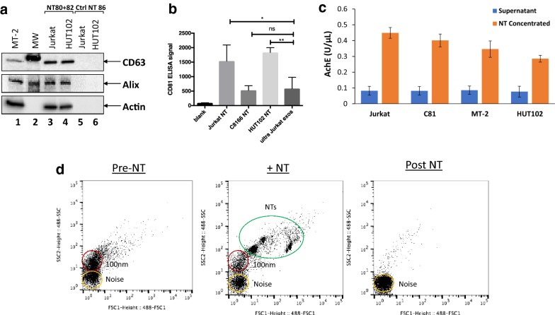
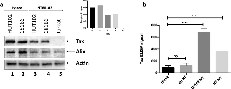
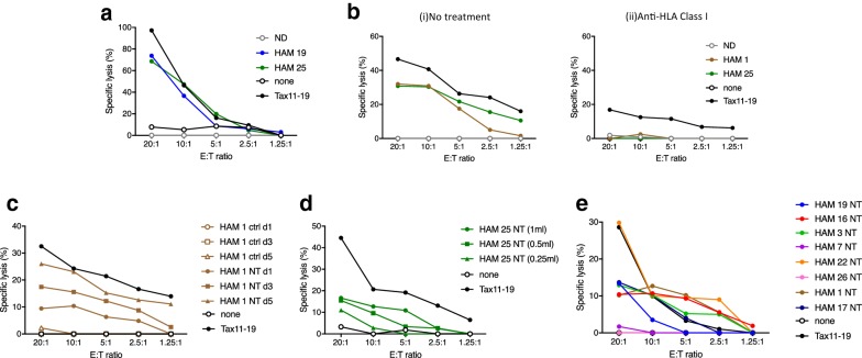
Results: Exosomes that were positive for HTLV-1 Tax by Western blot were isolated from HAM/TSP patient PBMCs (25/36) in ex vivo cultures by trapping exosomes from culture supernatants. HTLV-1 seronegative PBMCs did not have exosomes with Tax (0/12), (Fisher exact test, p = 0.0001). We were able to observe HAM/TSP patient CSF (12/20) containing Tax+ exosomes but not in HTLV-1 seronegative MS donors (0/5), despite the absence of viral detection in the CSF supernatant (Fisher exact test p = 0.0391). Furthermore, exosomes cultivated from HAM/TSP PBMCs were capable of sensitizing target cells for HTLV-1 specific CTL lysis.

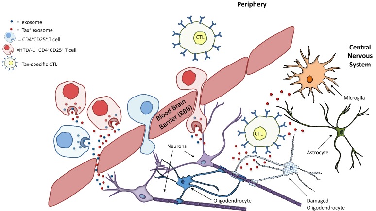
Conclusion: Cumulatively, these results show that there are HTLV-1 proteins present in exosomes found in virus-free CSF. HAM/TSP PBMCs, particularly CD4+CD25+ T cells, can excrete these exosomes containing HTLV-1 Tax and may be a source of the exosomes found in patient CSF. Importantly, these exosomes are capable of sensitizing an HTLV-1 specific immune response, suggesting that they may play a role in the immunopathology observed in HAM/TSP. Given the infiltration of HTLV-1 Tax-specific CTLs into the CNS of HAM/TSP patients, it is likely that exosomes may also contribute to the continuous activation and inflammation observed in HAM/TSP, and may suggest future targeted therapies in this disorder.

Keywords: Cell-free virus; Exosomes; HTLV-1 Tax; Nanotraps; Specific lysis; Spontaneous proliferation.


Figures

Fig. 1
Fig. 1
Nanotrapping exosomes from tissue culture supernatants. a CD63, Alix, and Actin levels from exosomes nanotrapped by either NT80 + 82 particles (lanes 3 and 4) or by NT86 particles (Ctrl NT 86; lanes 5 and 6) from Jurkat (HTLV-1 uninfected) and HUT102 (HTLV-1 infected) tissue culture supernatants were analyzed by Western blot. MT-2 whole cell extracts and molecular weight (MW) ladder are shown (lanes 1 and 2). b CD81 ELISA signals were measured from isolated exosomes as a measure of exosome concentration. Exosomes were nanotrapped (designated by NT) from HTLV-1 negative Jurkat, and HTLV-1 infected C8166 and HUT102 cells from 1 mL of starting tissue culture supernatant. The background is designated as blank. Ultracentrifuged Jurkat exosomes (ultra Jurkat exos) were also measured. Statistical analysis was performed by unpaired t-test. ns not significant; *p-value < 0.05; **p-value < 0.01 (n − 4). c Measurement of acetylcholinesterase (AchE) activity associated with exosome populations was performed. Tissue culture supernatant was measured for AchE activity from Jurkat, C8166 (C81), MT-2, and HUT102 cells both prior to nanotrapping (blue) and after addition of NT80 + 82 and subsequent exosome isolation (orange). d Tissue culture supernatants from HTLV-1 infected cells were analyzed by specialized flow cytometry for NanoFACS. Left panel represents a representative sample prior to addition of Nanotrap® particle (Pre-NT), middle panel represents sample with addition of Nanotrap® particle (+NT) and right panel represents sample after Nanotrap® particle and exosomes have been removed (Post NT). NTs are shown circled in green, while 100 nm vesicles are circled in red, and noise is circled in yellow
Fig. 2
Fig. 2
HTLV-1 infected cell lines have exosomes containing HTLV-1 Tax protein. a Western blots for levels of HTLV-1 Tax, Alix and β-actin (Actin) proteins were performed from cell lysates and NT80 + 82 nanotrapped exosomes from HUT102 and C8166 (C81) cells. Jurkat cell supernatants were also nanotrapped with NT80 + 82 particles and run alongside infected cell NT pulldowns. Tax levels detected were normalized to the β-actin signal for each sample and quantified in the upper right inset. b HTLV-1 Tax was measured by electrochemical ELISA. Nanotrapped (NT80 + 82) exosomes from Jurkat (Ju), C8166 (C81), and HUT102 (HT) cells were freeze-thawed and then measured for HTLV-1 Tax reactivity. Statistical significance was determined by unpaired t-test. ****p-value < 0.0001 (n = 4)
Fig. 3
Fig. 3
Exosome production from cultured PBMCs. a Normal donor (ND) or HAM/TSP (HAM) PBMCs were cultured in exosome free media and measured over 2–5 days for 3H-thymidine uptake in counts per minute (CPM). NDs were left unstimulated by IL-2 or CD3 (ND unstim). b Carboxyfluorescein succinimidyl ester (CFSE) signal by HAM/TSP PBMCs was measured after 5 days in culture. One representative graph is pictured. c ELISAs were performed over 5 days for CD81 from nanotrapped (NT80 + 82) exosomes from ND and HAM PBMCs. ND PBMCs were maintained with IL-2 to induce proliferation. d Exosomes were nanotrapped (NT80 + 82) after 5 days culture of HAM or ND PBMCs (n = 5), followed by ELISA for CD81. ND PBMCs were maintained with IL-2 to induce proliferation. e A CD81 ELISA was performed from nanotrapped (NT80 + 82) exosomes isolated from HAM 1 and HAM 17 patient samples over 5 days in culture. f Vesicles isolated from HAM PBMC (HAM 4, 19, 21, 25, and 30) short-term cultures by NT pulldown were measured for AchE activity. g ND PBMCs were sorted immediately after thawing (IL-2 only) or after activation for 1 day with anti-CD3 100 ng/mL and 100 IU/mL IL-2 (activated). h HAM/TSP PBMCs were cultured for 24 h prior to FACS sorting into CD4+CD25, CD4+CD25+, CD8+CD25, and CD8+CD25 T cells (n = 4). Statistical analysis was performed by unpaired t-test. *p-value < 0.05; **p-value < 0.01
Fig. 4
Fig. 4
Cultured HAM/TSP PBMCs produce exosomes containing HTLV-1 Tax. a HAM 1 and HAM 17 PBMCs were cultured over 5 days and were assessed for HTLV-1 Tax content from isolated exosomes by electrochemical ELISA. b A representative Western blot of HTLV-1 Tax and Actin from nanotrapped (NT80 + 82) exosomes of 7 HAM/TSP patient PBMCs after 5 days culture. Tax compared to Actin (β-actin) levels was quantified for the Western blot and shown in the upper right insert. NS nonspecific band. c An additional Western blot of HTLV-1 Tax, CD63 (two forms: glycosylated (CD63-Gly) and unmodified), and Actin from nanotrapped (NT80 + 82) exosomes of 4 ND and 4 HAM/TSP patient PBMCs after 5 days culture was performed. ND PBMCs were cultured with 100 IU/mL IL-2 while HAM/TSP PBMCs received no exogenous cytokines. Samples were run on a 4–20% Tris–glycine gel, followed by overnight wet transfer to PVDF membranes. C81 whole cell extract (WCE) was utilized as Tax positive control. NS nonspecific band, MW  molecular weight ladder
Fig. 5
Fig. 5
Detection of exosomes containing HTLV-1 Tax in CSF of HAM/TSP patients. a Cell-free CSF samples from 8 HAM/TSP patients and 2 MS patients were nanotrapped (NT80 + 82) and analyzed by ELISA for CD81 signal to determine levels of isolated exosomes. b Four HAM/TSP (HAM) and 5 multiple sclerosis (MS) patient CSF samples were nanotrapped (NT80 + 82), followed by Western blot analysis for Tax and Actin. C8166 whole cell extract (C81 WCE, lane 1) was added as a positive control. Tax normalized to Actin (β-actin) signal in the upper Western blot panel was quantified and is shown in the upper right inset. c Twelve HAM/TSP CSF samples were incubated with NT80 + 82 particles, followed by Western blot analysis for Tax and Actin levels. d Presence of HTLV-1 virus was assessed by analysis for the provirus. Pelleted cells and corresponding cell-free supernatants from HAM/TSP CSF samples were probed for HTLV-1 Tax by digital droplet PCR (ddPCR)
Fig. 6
Fig. 6
HAM/TSP exosomes can sensitize targets to Ag-specific responses. a Cytotoxic T lymphocyte (CTL) lysis was analyzed after the addition of HAM/TSP or ND PBMC-derived exosomes or Tax11-19 peptide. b HAM/TSP or ND PBMC-derived exosomes or Tax11-19 peptide pulsed targets were co-cultured with anti-HLA Class I ab (MCA81EL 5 μg/mL) and CTL target lysis was analyzed. c HAM/TSP PBMC supernatants were either nanotrapped with NT80 + 82 (NT) or with a control nanotrap (NT86) unable to isolate exosomes (ctrl). Supernatants collected after day 1, day 3, or day 5 of culture were used for trapping. d Various amounts (1 mL, 500, and 250 μL) of 5 days HAM 25 PBMC tissue culture supernatants were used as the starting material for nanotrapping by NT80 + 82 particles (NT), followed by CTL assay. e Eight representative HAM/TSP patient PBMC-derived exosomes were utilized for additional analysis of CTL lysis by CTL assay
Fig. 7
Fig. 7
Hypothesized contribution of exosomes to HAM/TSP immunopathogenesis. HTLV-1 infected PBMCs, especially HTLV-1 infected CD4+CD25+ T cells, produce exosomes containing HTLV-1 Tax. These cells are present in the periphery and produce Tax+ exosomes in the periphery that can potentially cross the blood brain barrier (BBB). However, these infected T cells are also capable of crossing the BBB and producing exosomes in the CNS. Both scenarios may explain the presence of Tax+ exosomes in HAM/TSP patient CSF. These Tax+ exosomes can then be taken up by CNS resident cells, including microglia, oligodendrocytes, astrocytes, etc. and become targets for lysis by HTLV-1 Tax-specific CTLs which cross the BBB. The resulting destruction perpetuates the neuroinflammation observed in HAM/TSP

References

    1. Koyanagi Y, Itoyama Y, Nakamura N, Takamatsu K, Kira J, Iwamasa T, et al. In vivo infection of human T-cell leukemia virus type I in non-T cells. Virology. 1993;196(1):25–33. doi: 10.1006/viro.1993.1451. - DOI - PubMed
    1. Satou Y, Matsuoka M. HTLV-1 and the host immune system: how the virus disrupts immune regulation, leading to HTLV-1 associated diseases. J Clin Exp Hematopathol. 2010;50(1):1–8. doi: 10.3960/jslrt.50.1. - DOI - PubMed
    1. Proietti FA, Carneiro-Proietti AB, Catalan-Soares BC, Murphy EL. Global epidemiology of HTLV-I infection and associated diseases. Oncogene. 2005;24(39):6058–6068. doi: 10.1038/sj.onc.1208968. - DOI - PubMed
    1. Evangelou IE, Massoud R, Jacobson S. HTLV-I-associated myelopathy/tropical spastic paraparesis: semiautomatic quantification of spinal cord atrophy from 3-dimensional MR images. J Neuroimaging. 2014;24(1):74–78. doi: 10.1111/j.1552-6569.2011.00648.x. - DOI - PMC - PubMed
    1. Yamano Y, Takenouchi N, Li HC, Tomaru U, Yao K, Grant CW, et al. Virus-induced dysfunction of CD4+CD25+ T cells in patients with HTLV-I-associated neuroimmunological disease. J Clin Invest. 2005;115(5):1361–1368. doi: 10.1172/JCI23913. - DOI - PMC - PubMed