Comparative Study
. 2018;46(sup3):S434-S447.
doi: 10.1080/21691401.2018.1499660. Epub 2018 Aug 27.

Comparison of covalently and physically cross-linked collagen hydrogels on mediating vascular network formation for engineering adipose tissue

Affiliations
Comparative Study

Comparison of covalently and physically cross-linked collagen hydrogels on mediating vascular network formation for engineering adipose tissue

Chia-Hui Chuang et al. Artif Cells Nanomed Biotechnol. 2018.

Abstract

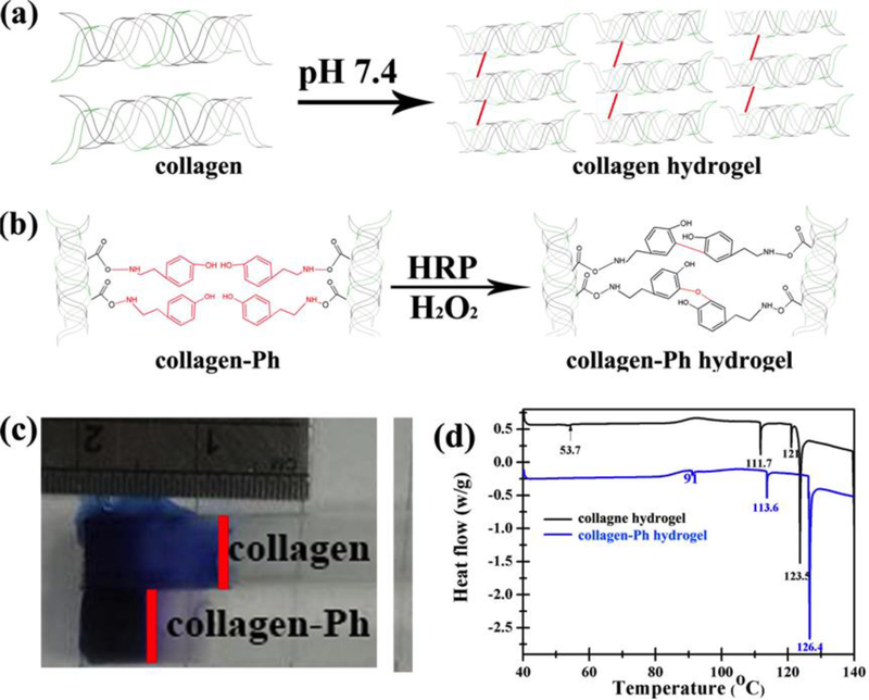
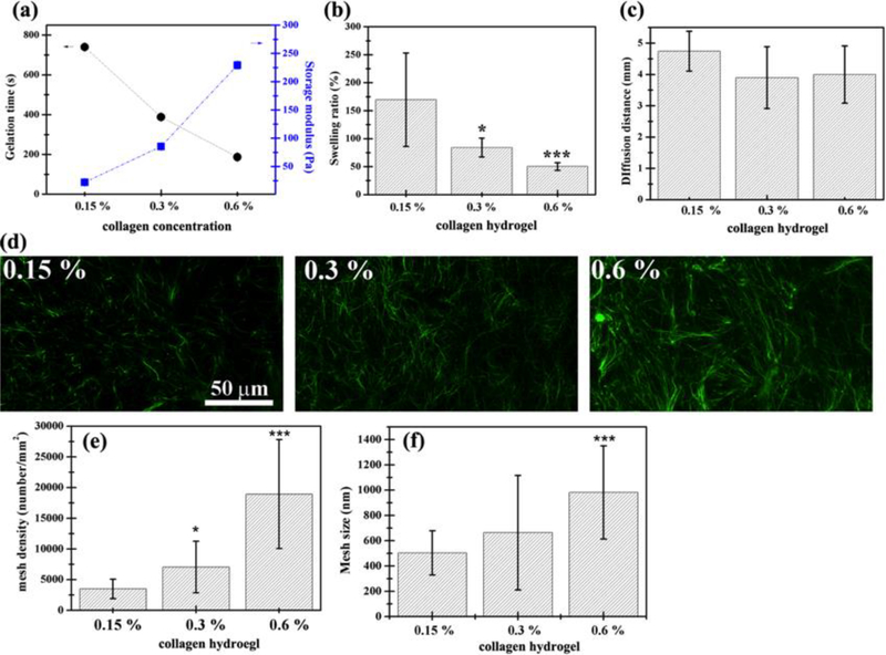
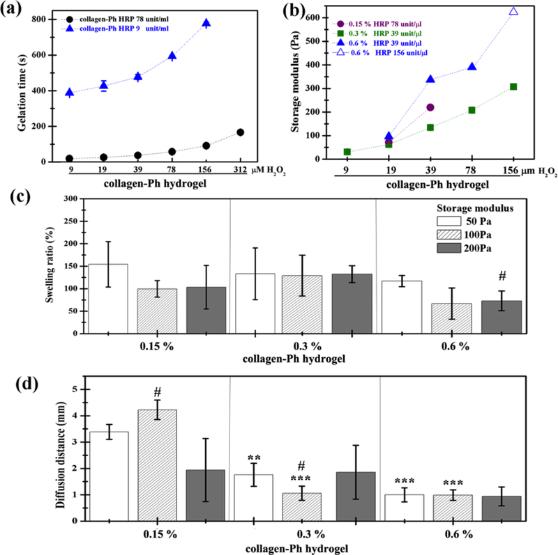
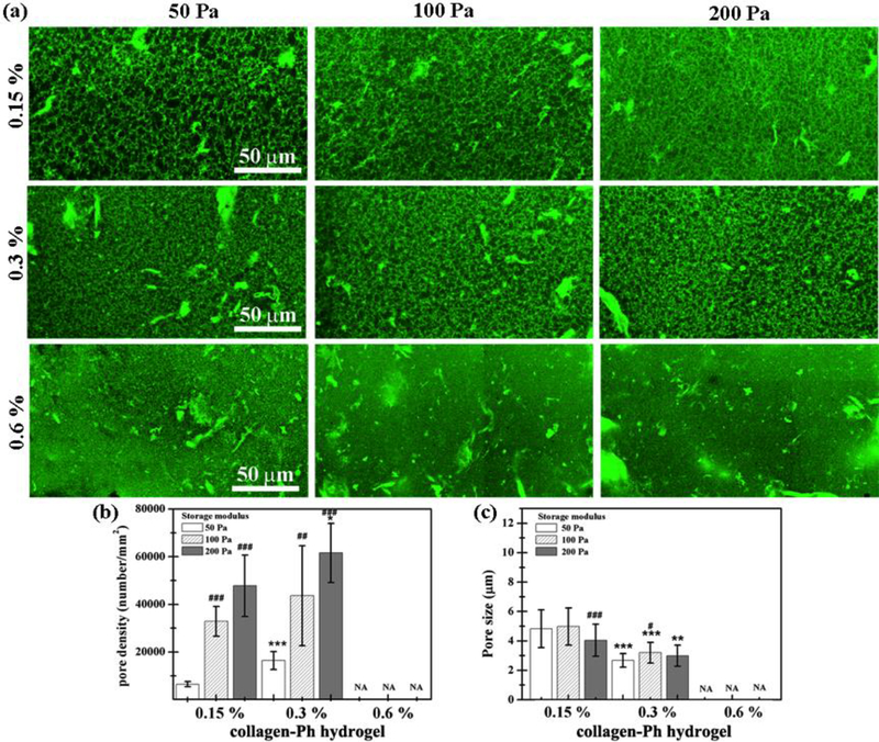
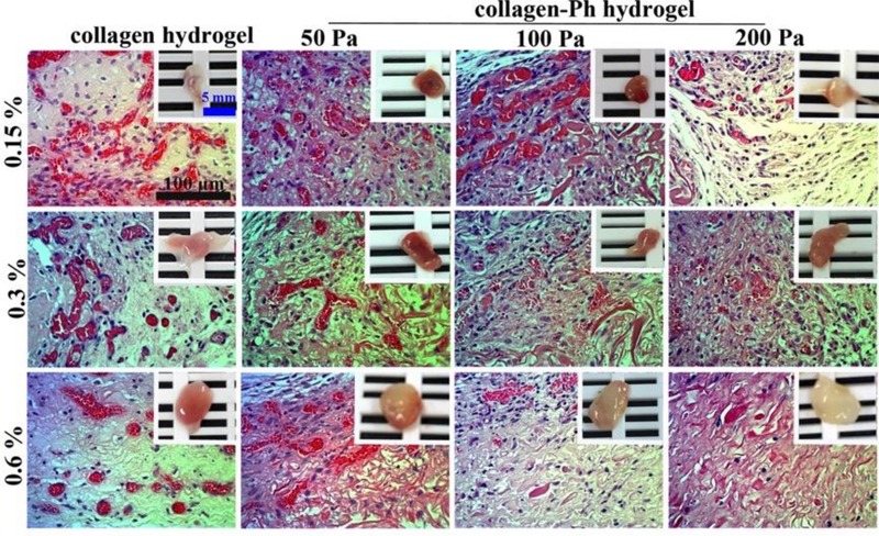
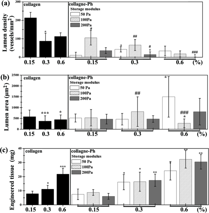
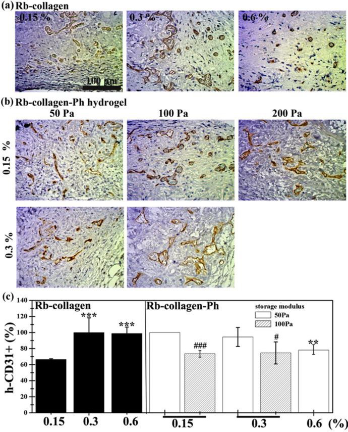
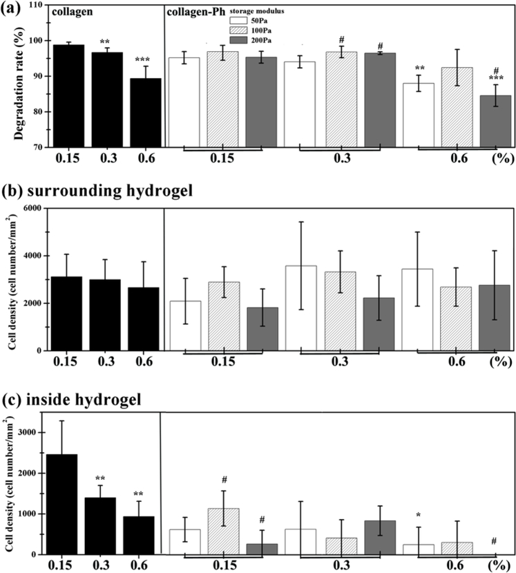
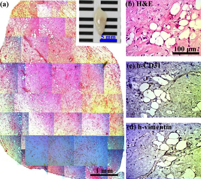
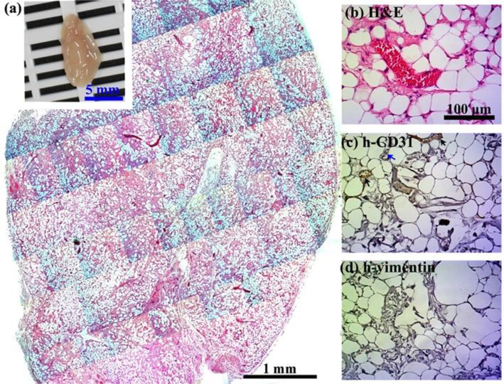
Timely tissue vascularization and integration of engineered tissues into a patient plays an important role in the successful translation of engineered tissues into clinically relevant therapies. To decrease the time needed to vascularize an engineered adipose tissue, suitable local microenvironments provided by hydrogels to support cell-based functional vascular network formation have been investigated. Using the same biomolecule in solution, two types of hydrogels can be obtained: a "physical hydrogel" which is thermal-induced self-assemble fibril initiation and growth, due to amino and carboxyl telopeptides on collagen chains, and a "chemical hydrogel" which results from the covalently cross-linking of the side chains induced by one step enzyme mediation in aqueous solution. In this paper, we compare the capability of engineering vascular network and large-sized vascularized adipose tissue in vivo in different types of collagen hydrogels, physical and chemical crosslinking. The relationships between vascular network formation and hydrogel properties for the two types of hydrogels are discussed. Finally, we successfully engineered a vascularized adipose tissue construct (∼877.6 adipocytes/mm2; 94% area of a construct) in the absence of exogenous cytokines in chemical covalently crosslinking cell-laden hydrogel. These results show manipulating the polymerized methods of a hydrogel could not only modulate vascular network formation, but also regenerate adipose tissue in vivo.

Keywords: Adipose tissue engineering vascular network; endothelial colony forming cells; hydrogel.


Figures

Figure 1.
Figure 1.
Formation of (a) physical cross-linked collagen hydrogels and (b) chemical cross-linked collagen-Ph hydrogels. The difference in (c) diffusion property and (d) energy of crosslinking bonds performed by differential scanning calorimetry on collagen and collagen-Ph hydrogels with same collagen concentration of 0.3 % and storage modulus of 100 Pa.
Figure 2.
Figure 2.
Physical properties of physical cross-linked collagen hydrogels vary with collagen concentration with limited tunability. (a) Gelation time decreased and shear storage modulus (G’) increased with increasing collagen concentration. (b) Swelling properties decreased with increasing collagen concentration. (c) Diffusion properties of 3D collagen hydrogel remains the same with increasing collagen concentration. (d-f) Mesh density increased linearly with collagen concentration as shown in (d) 3D confocal microscopy images. (e-f) Summarized data of 0.15, 0.3, and 0.6 % collagen hydrogels. P-values less than 0.1 and 0.001 were considered statistically significant and were labeled * and *** compared with the 0.15 % collagen hydrogel.
Figure 3.
Figure 3.
Tunable physical properties of chemical cross-linked collagen-Ph hydrogels varied with HRP, H2O2, and collagen-Ph concentration. (a) Gelation time was adjustable by HRP concentration. (b) Shear storage modulus (G’) increased with increasing collagen concentration and H2O2 concentration with a suitable amount of HRP. (c) Swelling properties remained constant with increasing collagen-Ph concentration or increasing G’. (d) Diffusion properties of collagen-Ph hydrogel as a function of collagen-Ph concentration and G’. Diffusion distances are larger than 3 mm for 0.15 % collagen-Ph hydrogels with G’=50 and 100 Pa, whereas others are below 2 mm. P-values less than 0.01 and 0.001 were considered statistically significant and were labeled ** and *** compared with the 0.15% collagen-Ph hydrogel with the same G’. P-values less than 0.1 were considered statistically significant and were labeled # compared with the collagen-Ph hydrogel with G’ of 50 Pa at the same concentration.
Figure 4.
Figure 4.
Collagen-Ph hydrogel microstructure as a function of collagen-Ph concentration and G’. (a) 3D confocal microscopy images and (b-c) summarized data for pore density and pore size are shown. P-values less than 0.01, 0.001 were considered statistically significant and were labeled ** and *** compared with the 0.15 % collagen-Ph hydrogel with the same G’. P-values less than 0.1, 0.01, 0.001 were considered statistically significant and were labeled #, ##, and ###, respectively compared with the collagen-Ph hydrogel with G’ of 50 Pa at the same concentration.
Figure 5.
Figure 5.
Crosslinking modulation of vascularized collagen constructs containing human ECFCs and MSCs collagen or collagen-Ph explanted after 7 days in vivo. H&E-stained images of vascularized collagen constructs with different cross-linking methods, G’, and protein concentrations, showing erythrocyte-filled lumens. The insets in the top panels show macroscopic views of the explants (scale bar 5 mm).
Figure 6.
Figure 6.
(a) Lumen density, (b) lumen area, and (c) weight of engineered cell-laden collagen (left) or collagen-Ph (right) tissue constructs were quantified. P-values less than 0.1, 0.01, 0.001 were considered statistically significant and were labeled *, **, and *** compared with the 0.15 % collagen-Ph hydrogel with the same G’. P-values less than 0.1, 0.01, 0.001 were considered statistically significant and were labeled #, ##, and ### compared with the collagen-Ph hydrogel with G’ of 50 Pa at the same concentration.
Figure 7.
Figure 7.
Crosslinking modulation of human ECFCs-MSC-mediated vascular network formation in collagen (left) or collagen-Ph (right) hydrogels after 7 days in vivo. Representative images of sections stained with human CD31+ ECFCs identified by immunohistochemistry shown in (a) collagen and (b) collagen-Ph hydrogels at various conditions. (c) The extent of human vascular network formation was quantified by counting erythrocyte-filled lumens, showing the percentage of the total blood vessels expressing human CD31. In collagen hydrogels, P-values less than 0.001 were considered statistically significant and were labeled *** compared with the 0.15% collagen hydrogel. In collagen-Ph hydrogels, P-values less than 0.01 were considered statistically significant and were labeled ** compared with the 0.15% collagen-Ph hydrogel with the same G’. P-values less than 0.1 and 0.001 were considered statistically significant and were labeled # and ### compared with the collagen-Ph hydrogel with G’ of 50 Pa at the same concentration.
Figure 8.
Figure 8.
Crosslinking modulation of biodegradation and host cell recruitment. Collagen and collagen-Ph solutions were subcutaneously injected into nude mice and polymerized to form collagen (left) and collagen-Ph (right) hydrogels. Constructs were evaluated after 7 days in vivo. (a) Modulation of in vivo degradation profiles on collagen hydrogels and collagen-Ph hydrogels with various amounts of collagen-Ph and G’. Quantification of total murine myeloid cells (b) surrounding host tissue and (c) inside the hydrogel constructs. In collagen hydrogels, P-values less than 0.01 and 0.001 were considered statistically significant and were labeled ## and ### compared with the 0.15% collagen hydrogel. In collagen-Ph hydrogels, P-values less than 0.1, 0.01, 0.001 were considered statistically significant and were labeled *, **, and *** compared with the 0.15% collagen-Ph hydrogel with the same G’. P-values less than 0.1 were considered statistically significant and were labeled # compared with the collagen-Ph hydrogel with G’ of 50 Pa at the same concentration.
Figure 9.
Figure 9.
Crosslinking modulation of vascularized adipose tissue graft in cell-laden collagen hydrogels. 0.6 % Collagen pre-polymer solutions with ECFCs and MSCs in the absence of any exogenous cytokines were subcutaneously injected into nude mice and formed cell-laden collagen hydrogels. Constructs were evaluated after 1 month in vivo. (a) The macroscopic view (inset, scale bar 5 mm) and representative H&E-stained section of entire cell-laden collagen constructs are shown (scale bar 1 mm). (b-d) High magnification of selected regions show some murine adipocytes (human-vimentin-negative) with murine vessels (human CD31-negative, blue arrows) carrying erythrocytes.
Figure 10.
Figure 10.
Crosslinking modulation of vascularized adipose tissue graft in collagen-Ph hydrogels. 0.6% Collagen-Ph pre-polymer solutions with ECFCs and MSCs in the absence of any exogenous cytokines were subcutaneously injected into nude mice and formed cell-laden collagen-Ph hydrogels under suitable concentrations of HRP and H2O2. Constructs were evaluated after 1 month in vivo. (a) The macroscopic view (inset, scale bar 5 mm) and representative H&E-stained section of entire cell-laden collagen-Ph constructs are shown (scale bar 1 mm). Adipose tissue covers entire construct. (b-d) High magnification of selected regions shows some murine adipocytes (human vimentin-negative) with some human vessels (human CD31-positive, black arrows)/murine vessels (human CD31-negative, blue arrows) carrying erythrocytes.

References

    1. Van Nieuwenhove I, Tytgat L, Ryx M, et al. Soft tissue fillers for adipose tissue regeneration: From hydrogel development toward clinical applications [Review]. Acta Biomater. 2017. November;63:37–49. doi: 10.1016/j.actbio.2017.09.026. PubMed PMID: ; English. - DOI - PubMed
    1. Choi JH, Gimble JM, Lee K, et al. Adipose Tissue Engineering for Soft Tissue Regeneration [Article]. Tissue Eng Part B-Rev. 2010. August;16(4):413–426. doi: 10.1089/ten.teb.2009.0544. PubMed PMID: ; English. - DOI - PMC - PubMed
    1. Coleman SR, Saboeiro AP. Fat grafting to the breast revisited: Safety and efficacy [Article; Proceedings Paper]. Plast Reconstr Surg. 2007. March;119(3):775–785. doi: 10.1097/01.prs.0000252001.59162.c9. PubMed PMID: ; English. - DOI - PubMed
    1. Smith P, Adams WP, Lipschitz AH, et al. Autologous human fat grafting: Effect of harvesting and preparation techniques on adipocyte graft survival [Article; Proceedings Paper]. Plast Reconstr Surg. 2006. May;117(6):1836–1844. doi: 10.1097/01.prs.0000218825.77014.78. PubMed PMID: ; English. - DOI - PubMed
    1. Rossi E, Gerges I, Tocchio A, et al. Biologically and mechanically driven design of an RGD-mimetic macroporous foam for adipose tissue engineering applications [Article]. Biomaterials. 2016. October;104:65–77. doi: 10.1016/j.biomaterials.2016.07.004. PubMed PMID: ; English. - DOI - PubMed

Publication types