. 2018 Aug 10:12:544.
doi: 10.3389/fnins.2018.00544. eCollection 2018.

C. elegans MANF Homolog Is Necessary for the Protection of Dopaminergic Neurons and ER Unfolded Protein Response

Affiliations

C. elegans MANF Homolog Is Necessary for the Protection of Dopaminergic Neurons and ER Unfolded Protein Response

Cory Richman et al. Front Neurosci. .

Abstract

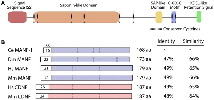
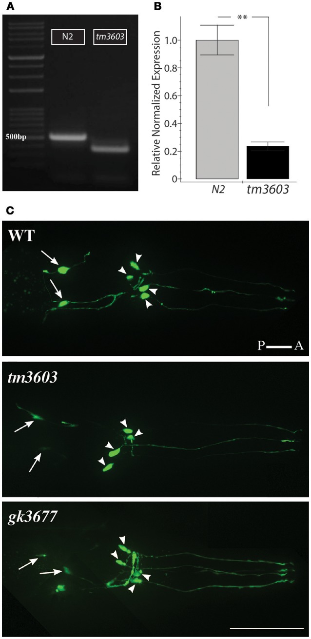
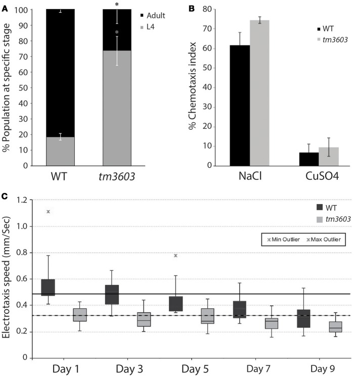
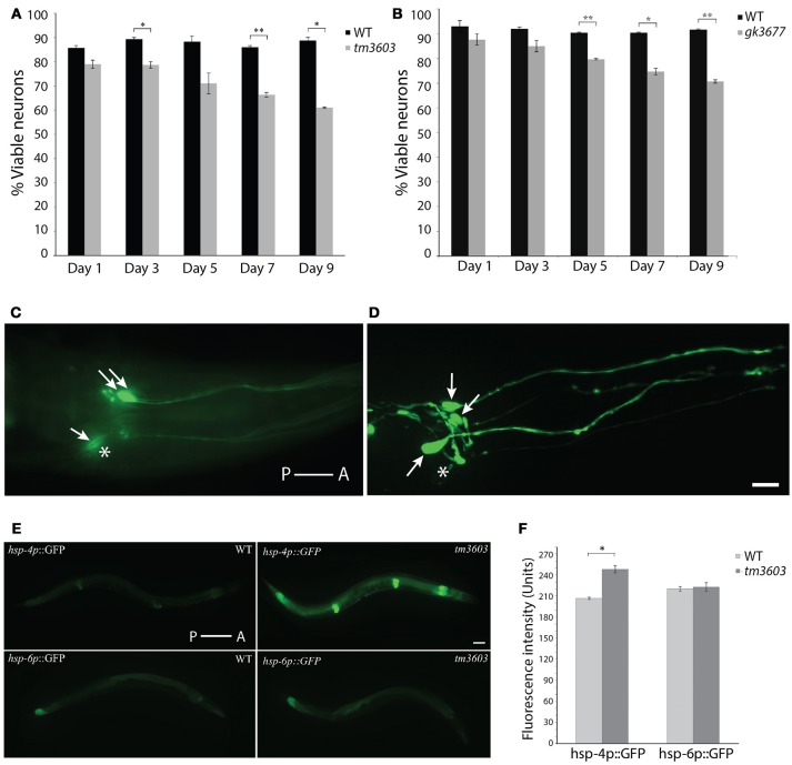
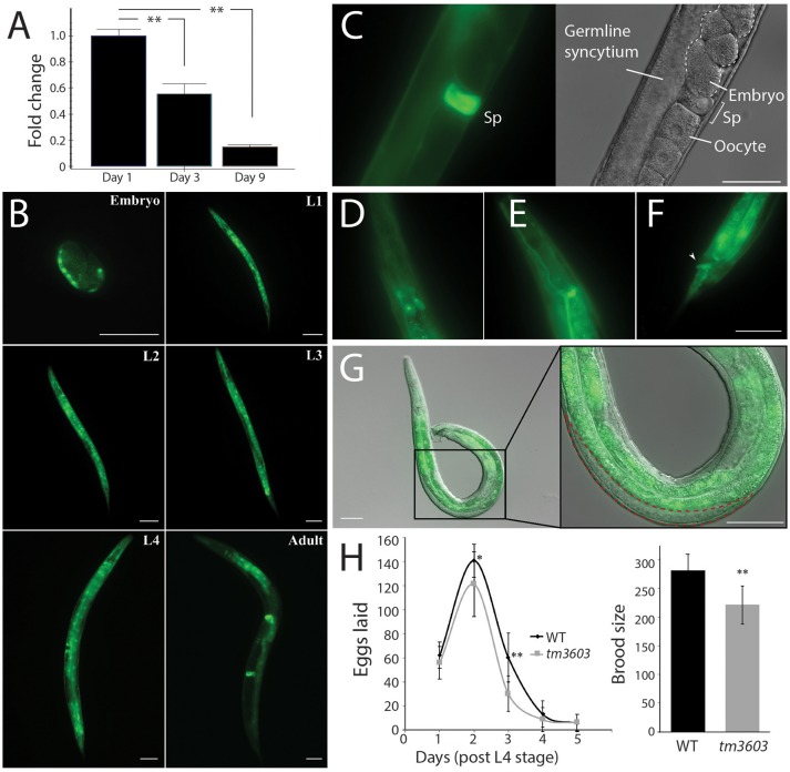
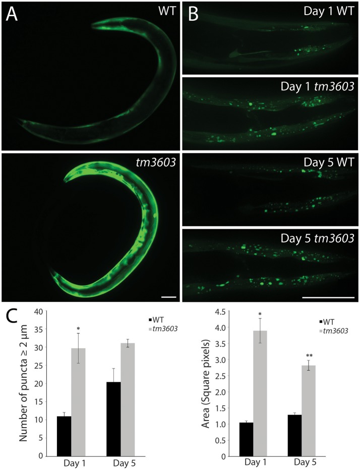
Neurotrophic factors (NTFs) are important for the development, function, and survival of neurons in the mammalian system. Mesencephalic astrocyte-derived neurotrophic factor (MANF) and cerebral dopamine neurotrophic factor (CDNF) are two recently identified members of a novel family of NTFs in vertebrates that function to protect dopaminergic neurons. Although these genes are conserved across eukaryotes, their mechanism of neuroprotection is not fully understood. Sequence searches for MANF/CDNF homologs in invertebrates have identified a single ortholog that is most related to MANF. Here we report the in vivo characterization of the MANF gene, manf-1, in the nematode Caenorhabditis elegans. We found that manf-1 mutants have an accelerated, age-dependent decline in the survival of dopaminergic neurons. The animals also show increased endoplasmic reticulum (ER) stress, as revealed by reporter gene expression analysis of hsp-4, an ER chaperone BiP/GRP78 homolog, suggesting that a failure to regulate the ER unfolded protein response (ER-UPR) may be a contributing factor to dopaminergic neurodegeneration. Expression studies of manf-1 revealed that the gene is broadly expressed in a pattern that matches closely with hsp-4. Consistent with the requirements of manf-1 in the ER-UPR, we found that aggregates of α-Synuclein, a major constituent of Lewy bodies, were significantly increased in body wall muscles of manf-1 mutant animals. Overall, our work demonstrates the important role of manf-1 in dopaminergic neuronal survival and the maintenance of ER homeostasis in C. elegans.

Keywords: C. elegans; CDNF; ER stress; MANF; Parkinson's disease; dopamine; manf-1; neurodegeneration.


Figures

Figure 1
Figure 1
MANF and CDNF homologs in selected organisms. (A) Protein domains and structure of the C. elegans manf-1 protein. (B) Schematic representation of MANF and CDNF proteins from Caenorhabditis elegans (Ce), Drosophila melanogaster (Dm), Homo sapiens (Hs) and Mus musculus (Mm) with percent identity and similarity indicated relative to C. elegans manf-1. Conserved domains are aligned and signal sequences (SS) are depicted with sizes, all presented to scale. GenBank accession numbers for amino acid sequences are described in Figure S1.
Figure 2
Figure 2
Characterization of manf-1 alleles. (A) manf-1 cDNA from wildtype (N2) and mutant (tm3603). 500 bp DNA ladder is marked for size comparison. (B) qRT-PCR analysis of manf-1 in 1 day old adults. Results are means of experiments performed in triplicate ± SEMs, **p < 0.01. (C) Visualization of dopaminergic neurons in the head of wildtype (WT), tm3603 and gk3677 mutant alleles. The posterior pair of ADEs is indicated by arrows and four anterior CEPs are indicated by arrowheads. Scale bar = 50 μm.
Figure 3
Figure 3
Chemosensory and life traits of manf-1(tm3603) animals. (A) Growth delay represented by percentage of animals reaching adulthood by 54 h after plating synchronized L1 larvae. Experiment performed in duplicate ± SEM, *p = 0.0144. (B) Results of chemotaxis assays for NaCl (chemo-attractant) and CuSO4 (chemo-repellent). Means are derived from four batches ± SEMs, demonstrating a functional and comparable chemotactic response between mutant and wildtype animals. (C) Box plot of the electrotaxis response of tm3603 and wildtype (N2) controls on 1, 3, 5, 7, and 9 days of adulthood. The horizontal line inside each box marks the median, with upper and lower ends of boxes representing 25 and 75th quartile of data samples. Each vertical line with caps shows the spread of data. Stars indicate outliers. The speed of animals declines with age resulting in a 37% reduction in wildtype by day-9 and 30% in tm3603 over the same period. The solid and dotted black horizontal lines serve as references for the median speeds of day-1 wildtype and tm3603 animals, respectively. p < 0.01 for all days compared.
Figure 4
Figure 4
Dopaminergic neuron and ER stress phenotypes of manf-1 mutants. Quantification of dopaminergic neuronal defects in day-1 to day-9 old adults of tm3603 (A) and gk3677 (B) mutant animals. In each case wildtype (WT) animals were used as controls. Experiments performed in duplicate ± SEMs, *p < 0.05, **p < 0.01. (C,D) Nomarski fluorescence (C) and Confocal (D) images of dopaminergic neurons in two different tm3603 day-7 old animals. Arrows mark neuronal cell bodies that are visible and stars placed next to those that are faint and/or undetectable. Scale bar = 25 μm. (E) Differential induction of ER and mitochondrial stress. Whole animal ER (hsp-4p::GFP) and mitochondrial (hsp-6p::GFP) reporters visualized in wildtype and tm3603 animals; worms with average fluorescence shown. Scale bar = 50 μm. (F) The average GFP pixel intensity values quantified via whole animal analysis, performed in triplicate ± SEMs, *p < 0.05. Anterior (A) and posterior (P) orientations in (C–E) are indicated.
Figure 5
Figure 5
Expression profile of manf-1 and fecundity defects in manf-1 mutants. (A) qRT-PCR data showing a decline in endogenous manf-1 transcript levels with age in wildtype animals. Results are means of experiments performed in duplicate ± SEMs, normalized to day-1 adult manf-1 expression levels, **p < 0.01. (B) GFP fluorescence in manf-1p::GFP animals at different stages of development (embryo, larval L1 to L4, and adult). (C) Bright fluorescence within the spermatheca (Sp) of adults. (D–F) Expression within the excretory system; excretory gland cells (D), canal (E), and anus (F) indicated by arrowhead. (G) Gonad of an adult manf-1p::GFP transgenic male showing a lack of fluorescence in the region containing sperm (indicated by red dotted line). (H) Frequency of egg laying and brood sizes in wildtype and tm3603 animals. Means ± SDs are plotted. *p < 0.05, ** < 0.01. Scale bar for all images = 50 μm.
Figure 6
Figure 6
manf-1 mutants show enhanced aggregation of α-Synuclein. (A) Expression analysis of human SNCA::YFP gene within the body wall muscles of wildtype (N2) and tm3603 animals. (B) Maximum projection of Z-stack images at days 1 and 5 of adulthood showing YFP fluorescent puncta. (C) Number of puncta of diameter 2 μm and above determined by manual counting and automated analysis of total pixel area after thresholding. Experiments performed in triplicate ± SEMs, *p < 0.05, ** < 0.01. Scale bar = 50 μm.

References

    1. Airavaara M., Shen H., Kuo C. C., Peränen J., Saarma M., Hoffer B., et al. . (2009). Mesencephalic astrocyte-derived neurotrophic factor reduces ischemic brain injury and promotes behavioral recovery in rats. J. Comp. Neurol. 515, 116–124. 10.1002/cne.22039 - DOI - PMC - PubMed
    1. Apostolou A., Shen Y., Liang Y., Luo J., Fang S. (2008). Armet, a UPR-upregulated protein, inhibits cell proliferation and ER stress-induced cell death. Exp. Cell Res. 314, 2454–2467. 10.1016/j.yexcr.2008.05.001 - DOI - PMC - PubMed
    1. Bai M., Vozdek R., Hnízda A., Jiang C., Wang B., Kuchar L., et al. . (2018). Conserved roles of C. elegans and human MANFs in sulfatide binding and cytoprotection. Nat. Commun. 9:897. 10.1038/s41467-018-03355-0 - DOI - PMC - PubMed
    1. Barbacid M. (1995). Neurotrophic factors and their receptors. Curr. Opin. Cell Biol. 7, 148–155. 10.1016/0955-0674(95)80022-0 - DOI - PubMed
    1. Bargmann C. I. (2006). Chemosensation in C. elegans, in The, C. elegans Research Community, ed WormBook. (WormBook; ), 1–29. Available online at: http://www.wormbook.org - PMC - PubMed

LinkOut - more resources