. 2018 Aug 10:9:1856.
doi: 10.3389/fimmu.2018.01856. eCollection 2018.

Allergic Asthma Favors Brucella Growth in the Lungs of Infected Mice

Affiliations

Allergic Asthma Favors Brucella Growth in the Lungs of Infected Mice

Arnaud Machelart et al. Front Immunol. .

Abstract

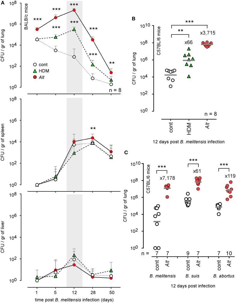
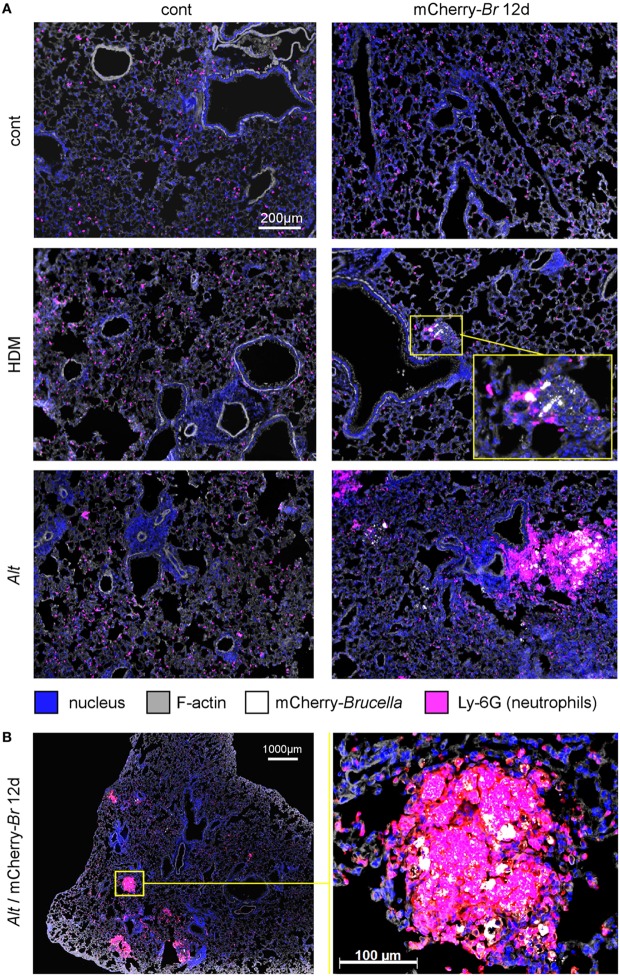
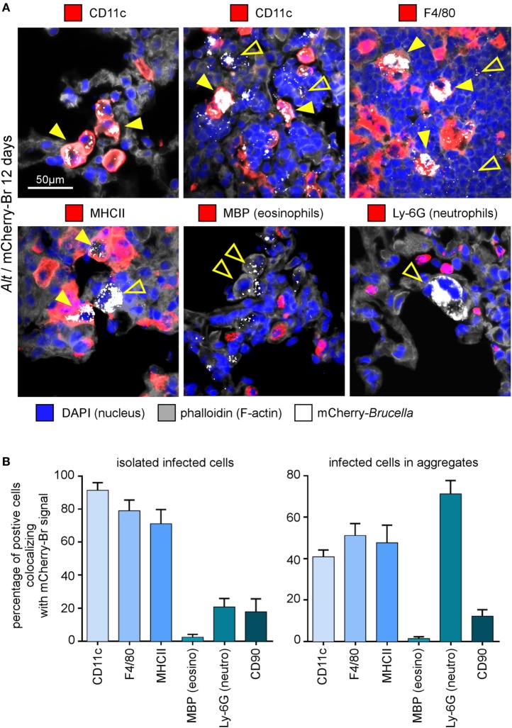
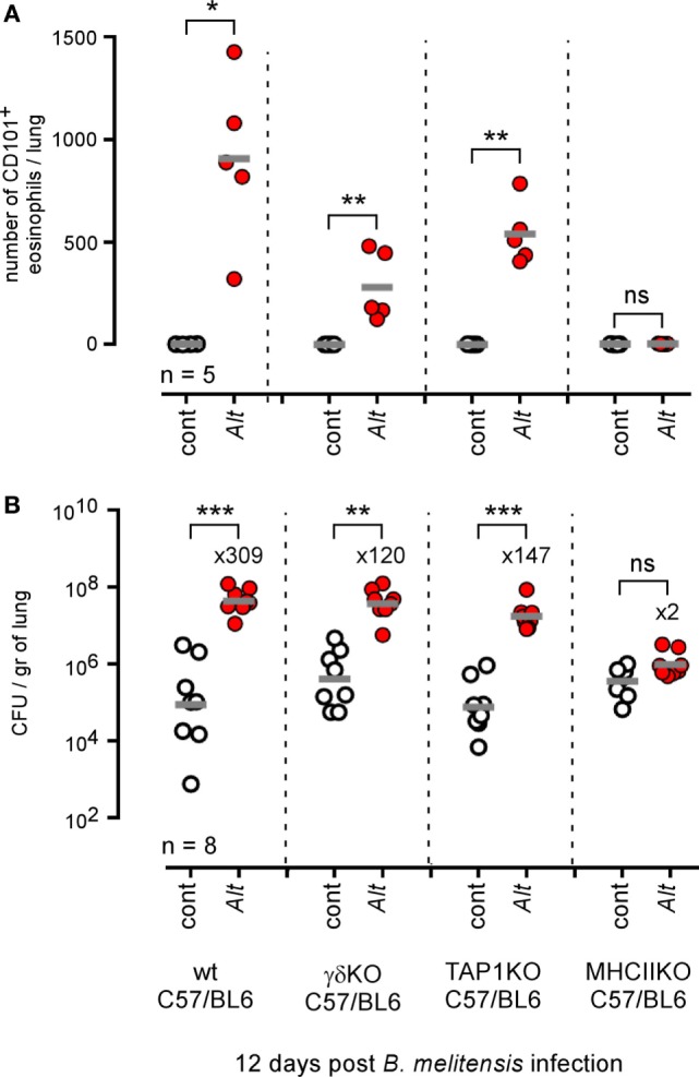
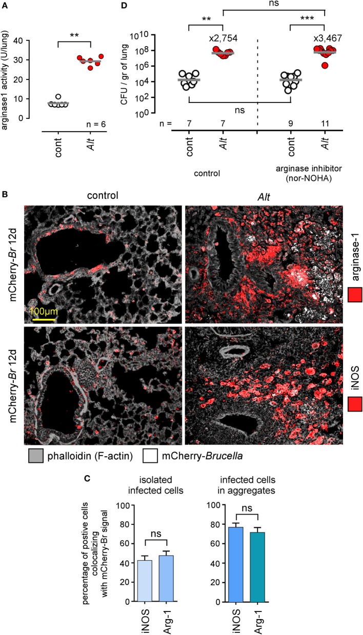
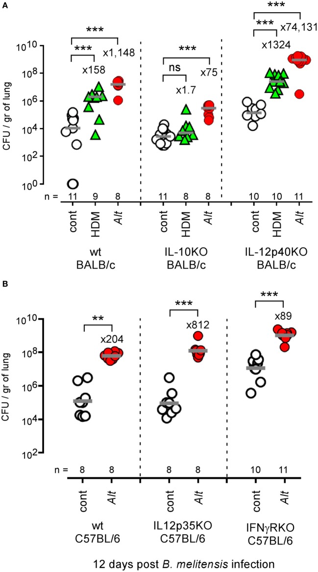
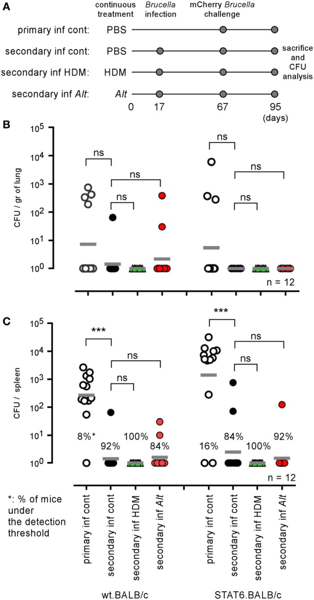
Allergic asthma is a chronic Th2 inflammatory disease of the lower airways affecting a growing number of people worldwide. The impact of infections and microbiota composition on allergic asthma has been investigated frequently. Until now, however, there have been few attempts to investigate the impact of asthma on the control of infectious microorganisms and the underlying mechanisms. In this work, we characterize the consequences of allergic asthma on intranasal (i.n.) infection by Brucella bacteria in mice. We observed that i.n. sensitization with extracts of the house dust mite Dermatophagoides farinae or the mold Alternaria alternata (Alt) significantly increased the number of Brucella melitensis, Brucella suis, and Brucella abortus in the lungs of infected mice. Microscopic analysis showed dense aggregates of infected cells composed mainly of alveolar macrophages (CD11c+ F4/80+ MHCII+) surrounded by neutrophils (Ly-6G+). Asthma-induced Brucella susceptibility appears to be dependent on CD4+ T cells, the IL-4/STAT6 signaling pathway and IL-10, and is maintained in IL-12- and IFN-γR-deficient mice. The effects of the Alt sensitization protocol were also tested on Streptococcus pneumoniae and Mycobacterium tuberculosis pulmonary infections. Surprisingly, we observed that Alt sensitization strongly increases the survival of S. pneumoniae infected mice by a T cell and STAT6 independent signaling pathway. In contrast, the course of M. tuberculosis infection is not affected in the lungs of sensitized mice. Our work demonstrates that the impact of the same allergic sensitization protocol can be neutral, negative, or positive with regard to the resistance of mice to bacterial infection, depending on the bacterial species.

Keywords: Brucella melitensis; Mycobacterium tuberculosis; Streptococcus pneumoniae; allergic asthma; brucellosis; infection.


Figures

Figure 1
Figure 1
Impact of allergic asthma sensitization on the course of Brucella infection in wild-type mice. Wild-type BALB/c and C57BL/6 mice received repeated i.n. administration of phosphate-buffered saline, HDM, or Alt before i.n. infection with 2 × 104 CFU of mCherry-Brucella melitensis (A,B) or 2 × 103 CFU of B. melitensis, B. abortus, or Brucella suis (C), as indicated in the figure and in the Section “Materials and Methods.” The mice were sacrificed at the selected time post infection. The data represent the number of CFU/gr of lung, spleen, and liver. n denotes the number of mice used for each lineage. These results are representative of at least two independent experiments. **p < 0.01, ***p < 0.001.
Figure 2
Figure 2
Microscopic analysis of lungs from control, HDM and Alt sensitized BALB/c mice. Wild-type BALB/c mice received repeated i.n. administration of phosphate-buffered saline (PBS) or Alt before i.n. inoculation of PBS or 2 × 104 CFU of mCherry-Brucella melitensis. The mice were sacrificed at 12 days post infection. The lungs were harvested and fixed. Frozen sections were examined by immunohistofluorescence for bacteria (mCherry signal) and Ly-6G-expressing cells. (A) Comparison of control (cont), HDM, and Alt sensitized mice, infected or not. (B) High magnification representative view from Alt sensitized infected mice. The panels are color-coded by Ag as indicated. The data are representative of two independent experiments.
Figure 3
Figure 3
Cell surface phenotype of Brucella melitensis infected cells in asthmatic lungs. Wild-type BALB/c mice received repeated i.n. administration of Alt before i.n. infection with 2 × 104 CFU of mCherry-B. melitensis. The mice were sacrificed at 12 days post infection. The lungs were harvested and fixed. Frozen sections were examined by immunohistofluorescence for mCherry (Brucella), CD11c, F4/80, MHCII, major basic protein (MBP), Ly-6G, and CD90 signals. The panels are color-coded by antigen as indicated. (A) High magnification representative view of infected cells in the lungs. (B) The data represent the percentage of mCherry-Br signal that co-localizes with CD11c, F4/80, MHCII, MBP, Ly-6G, and CD90 markers. When >12 infected cells are observed in the same observation field (approximately 700 μm × 500 μm), they are considered as “aggregated,” and when <12 are observed, they are considered as “isolated.” At least 200 infected cells from three different infected mice were analyzed for each staining. These data are taken from two independent experiments.
Figure 4
Figure 4
Alveolar macrophages and neutrophils are both infected with Brucella melitensis in the lungs of Alt-sensitized BALB/c mice. Wild-type BALB/c mice received repeated i.n. administration of Alt before i.n. infection with 2 × 104 CFU of mCherry-B. melitensis. The mice were sacrificed at 12 days post infection. The lungs were harvested, fixed, and frozen sections were immunostained and examined by confocal microscopy. (A,B) Representative confocal images of single optical sections of mCherry-Br infected CD11c+ cells and Ly-6G+ cells stained with DAPI from Alt sensitized wild-type BALB/c mice. (C) Representative infected multinucleated giant cells stained with DAPI and phalloidin. The panels are color-coded for DAPI, phalloidin, the cell surface antigen examined or mCherry-Brucella. Scale bars = 10 µm, as indicated.
Figure 5
Figure 5
Impact of IL-4 and STAT6 deficiency on asthma-induced Brucella susceptibility in BALB/c mice. Wild-type, IL4−/− and STAT6−/− BALB/c mice received repeated i.n. administration of phosphate-buffered saline, HDM, or Alt before i.n. infection with 2 × 104 CFU of mCherry-Brucella melitensis. The mice were sacrificed at 12 days post infection. The data represent the number of CFU/gr of lung at 12 days post infection from each group. n denotes the number of mice used for each lineage. These results are representative of at least two independent experiments. ***p < 0.001.
Figure 6
Figure 6
Impact of T lymphocyte deficiency on Alt-induced Brucella susceptibility. Wild-type, γδTCR−/−, TAP1−/−, and MHCII−/− C57BL/6 mice received repeated i.n. administration of phosphate-buffered saline or Alt before i.n. infection with 2 × 104 CFU of mCherry-Brucella melitensis. The mice were sacrificed at 12 days post infection. (A) The data represent the number of eosinophils recruited (SIGLEC-F+ CD11c CD101+) × 105 per lung from each group as determined by flow cytometry. (B) The data represent the number of CFU/gr of lung at 12 days post infection from each group. Horizontal gray lines represent the medians. n denotes the number of mice used for each lineage. These results are representative of at least three independent experiments. *p < 0.05, **p < 0.01, ***p < 0.001.
Figure 7
Figure 7
Asthma-induced Brucella susceptibility is independent of arginase activity. Wild-type BALB/c mice received repeated i.n. administration of phosphate-buffered saline (PBS) (control) or Alt before i.n. administration of PBS or 2 × 104 CFU of mCherry-Brucella melitensis. The mice were sacrificed at 12 days post infection. The lungs were harvested and fixed. Frozen sections were examined by immunohistofluorescence for mCherry (Brucella), arginase-1 and iNOS signals. During the sensitization and infection process, the mice received repeated administration of PBS (control group) or the arginase inhibitor nor-NOHA. (A) The panel represents the arginase activity/lung homogenate from control and Alt sensitized wild-type BALB/c mice. (B) Representative view of mCherry+, arginase+, and iNOS+ cells in the lungs of control and Alt sensitized mice. The panels are color-coded by antigen as indicated. (C) The data represent the percentage of mCherry-Br signal that co-localizes with arginase-1 and iNOS staining. When >12 infected cells are observed in the same observation field (approximately 700 μm × 500 μm), they are considered as “aggregated,” and when <12 are observed, they are considered as “isolated.” At least 200 infected cells from three different infected mice were analyzed for each staining. (D) The panel represents the number of CFU/gr of lung at 12 days post infection from each group of mice. Horizontal gray lines represent the medians. n denotes the number of mice used for each lineage. These results are representative of at least two independent experiments. **p < 0.01, ***p < 0.001.
Figure 8
Figure 8
Impact of IL-10 and IL-12 deficiency on asthma-induced Brucella susceptibility. (A) Wild-type, IL-10−/−, and IL-12p40−/− BALB/c mice received repeated i.n. administration of phosphate-buffered saline (PBS) or Alt before i.n. infection with 2 × 104 CFU of mCherry-Brucella melitensis. The mice were sacrificed at 12 days post infection. The data represent the CFUs per gram of lung. (B) Wild-type, IL-12p35−/−, and IFN-γR−/− C57BL/6 mice received repeated i.n. administration of PBS or Alt before i.n. infection with 2 × 104 CFU of mCherry-B. melitensis. The mice were sacrificed at 12 days post infection. The data represent the CFU/gr of lung. Horizontal gray lines represent the medians. n denotes the number of mice used for each lineage. These results are representative of at least three independent experiments. **p < 0.01, ***p < 0.001.
Figure 9
Figure 9
Characterization of IL-10 producing cells in lungs from infected asthmatic mice. IL-10GFP transgenic C57BL/6 mice were sensitized with phosphate-buffered saline (PBS) (control group), HDM, or Alt before i.n. infection with 2 × 104 CFU of Brucella melitensis. The mice were sacrificed at 12 days post infection, and the spleen cells were analyzed by flow cytometry. (A) Cells from PBS, HDM, and Alt infected mice were analyzed for IL-10GFP, CD3, and CD4 expression. The figure shows representative dot plots from individual lungs in each group, as indicated in the figure. Numbers represent the percentage of IL-10GFP+ cells in 50,000 acquired events. (B) The data represent the number of IL-10GFP+ eosinophils, γδ+ T cells, B cells, CD4+ T cells, CD8+ T cells, and natural killer cells per 105 lung cells from control, HDM, and Alt infected mice. The data are representative of two independent experiments.
Figure 10
Figure 10
Influence of allergic asthma on the development of protective memory against Brucella melitensis. Wild-type and STAT-6−/− BALB/c mice were sensitized with phosphate-buffered saline (PBS), HDM, or Alt extracts, as described in the Section “Materials and Methods,” throughout the entire experiment. Primary and secondary groups received i.n. administration of PBS or 2 × 104 CFU of wild-type B. melitensis at 17 days, respectively. All groups were infected i.n. with 2 × 104 CFU of mCherry-B. melitensis at 67 days, were sacrificed at 95 days, and the spleens were harvested and the CFUs were counted. (A) Is a schematic representation of the protocol. (B,C) Represent the number of CFU/gr of lung (B) and spleen (C). Horizontal gray lines represent the medians. n denotes the number of mice used for each lineage. These results are representative of at least two independent experiments. ***p < 0.001.
Figure 11
Figure 11
Asthma sensitization increases resistance against intranasal Streptococcus pneumoniae infection. Wild-type and CD3−/− C57BL/6 (A,B) and wild-type and STAT-6−/− BALB/c mice (C) were sensitized with phosphate-buffered saline (cont) or Alt before i.n. infection with a lethal CFU dose (2 × 107 CFU) of S. pneumonia, as indicated in the legend of each graph. (A,C) The data shown are the percentage of mice surviving infection at a given time. (B) The data represent the number of CFU per lung or spleen, as indicated. n indicates the number of mice per group. These results are representative of at least two independent experiments.
Figure 12
Figure 12
Asthma sensitization does not affect the course of Mycobacterium tuberculosis infection in the lungs of mice. Wild-type BALB/c mice were sensitized with phosphate-buffered saline (cont) or Alt before aerosol infection with 102 CFU of M. tuberculosis. Mice were sacrificed at the indicated time post M. tuberculosis infection. The data show the CFU/lung. n indicates the number of mice per group. These results are representative of at least two independent experiments.

References

    1. Stein RA. Super-spreaders in infectious diseases. Int J Infect Dis (2011) 15:e510–3.10.1016/j.ijid.2010.06.020 - DOI - PMC - PubMed
    1. Masoli M, Fabian D, Holt S, Beasley R. The global burden of asthma: executive summary of the GINA dissemination committee report. Allergy (2004) 59:469–78.10.1111/j.1398-9995.2004.00526.x - DOI - PubMed
    1. Anandan C, Nurmatov U, Van Schayck OCP, Sheikh A. Is the prevalence of asthma declining? Systematic review of epidemiological studies. Allergy (2010) 65:152–67.10.1111/j.1398-9995.2009.02244.x - DOI - PubMed
    1. Fahy JV. Type 2 inflammation in asthma — present in most, absent in many. Nat Immunol (2015) 15:57–65.10.1038/nri3786 - DOI - PMC - PubMed
    1. Stiemsma LT, Reynolds LA, Turvey SE, Finlay BB. The hygiene hypothesis: current perspectives and future therapies. Immunotargets Ther (2015) 4:143–57.10.2147/ITT.S61528 - DOI - PMC - PubMed

Publication types

MeSH terms