Review
. 2018 Aug;26(3):136-144.
doi: 10.1177/1742271X18762250. Epub 2018 Feb 28.

Vascular ultrasound, the potential of integration of multiparametric ultrasound into routine clinical practice

Affiliations
Review

Vascular ultrasound, the potential of integration of multiparametric ultrasound into routine clinical practice

Vasileios Rafailidis et al. Ultrasound. 2018 Aug.

Abstract

Introduction: Ultrasound has traditionally been regarded as the first-line imaging modality for screening, diagnostic evaluation and monitoring treatment and disease progression of vascular pathology, including both the arterial and the venous branch of the vascular system. Albeit of its well-tolerated nature, wide availability and low cost, ultrasound above all, has the advantage of providing the clinician with clinically significant information related to both intraluminal irregularities and extravascular disease. Ultrasound has the potential not only to anatomically describe tissues but also to assess physiology by evaluating blood flow characteristics in real time.

Discussion: The already fundamental role of ultrasound has been even more expanded with the introduction of newer techniques like contrast-enhanced ultrasound, tissue-elastography and 3D ultrasound and the incorporation of research advances into clinical practice. The purpose of this review is to present and discuss some of the latest advances in the field of vascular ultrasound in attempt to illustrate how the adoption of multiparametric ultrasound into everyday clinical practice could address the patient's needs. Pathology and applications discussed include carotid and aortic disease, portal and peripheral venous abnormalities.

Conclusion: Widespread availability of modern technology in ultrasound devices has made the application of contrast-enhanced ultrasound, tissue elastography and 3D ultrasound feasible options, ready to contribute to the diagnostic performance of the ultrasonographic technique.

Keywords: Vascular; aneurysm; aorta; contrast-enhanced ultrasound; portal vein.


Figures

Figure 1.
Figure 1.
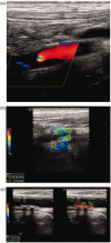
A 64-year-old male patient presenting with acute stroke and with an ipsilateral ulcerated ICA plaque. Long-axis colour Doppler image (a) shows a hypoechoic plaque causing severe stenosis (asterisk). Corresponding CEUS image (b) delineating a superficial ulceration (arrowhead) on this extremely stenotic plaque. Axial colour Doppler image (c) showing the stenotic ICA lumen surrounded by a hypoechoic plaque (asterisk). Corresponding CEUS image (d) confirming the presence of ulceration (arrowhead) (asterisk: ICA lumen; E: external carotid artery). CTA image (e) confirming CEUS findings, with contrast in the ulcerated plaque (arrowhead).
Figure 2.
Figure 2.
A 60-year-old male patient with an acute stroke. Ipsilateral carotid artery US confirmed the presence of severe ICA stenosis. Consecutive CEUS images (a, b, c) demonstrate the presence of moving microbubbles (arrowheads) within the initially hypoechoic part of the plaque; findings consistent with mild intraplaque neovascularization.
Figure 3.
Figure 3.
US and CEUS findings in a 57-year-old female patient with a right-sided stroke. Colour Doppler US (a) shows a hypoechoic plaque causing severe stenosis of the ipsilateral ICA (arrow). CEUS image immediately after the arrival of the microbubbles (b) demonstrates the plaque as uniformly hypoechoic and accurately delineates the plaque's border (arrowheads). Consecutive CEUS images (c, d) detected moving microbubbles (arrowheads) within the plaque, which gradually shows uniform enhancement (arrowheads); findings consistent with severe intraplaque neovascularization. A time-intensity curve (e) quantifies the plaque enhancement in comparison with the lumen enhancement. Parametric image (f) characterizing the plaque's enhancement based on the time of microbubble's arrival. It is assumed that the plaque enhances in a centrifugal pattern, probably due to the presence of adventitial neo-vessels (arrowheads pointing to the plaque).
Figure 4.
Figure 4.
Imaging findings in a 74-year-old male patient with pre-occlusive stenosis of the ICA. Colour Doppler US (a) raised suspicion of ICA occlusion. However, CEUS (b) readily identified a thread-like opacified lumen establishing the diagnosis of near-occlusion (arrow), which was also confirmed with CTA (arrowheads in c).
Figure 5.
Figure 5.
A 65-year-old male patient with stroke was diagnosed with an ipsilateral hypoechoic internal carotid artery plaque (colour Doppler US on a). Shear-wave elastography (b) has showed the plaque to be soft based on low elasticity values. A different 70-year-old asymptomatic patient was diagnosed with a heavily calcified internal carotid artery plaque which on shear-wave elastography was found to be hard based on high elasticity values.
Figure 6.
Figure 6.
An 80-year-old-man, following EVAR, with a persistent enlarged aneurysmal sac, with renal impairment undergoing surveillance with follow-up ultrasound. B-mode ultrasound (a) in an axial view demonstrating the stented aorta (arrow) and surrounding echogenic thrombus within the aneurysmal sac. Colour Doppler US (b) suggests a leak with colour Doppler signal (arrow) identified within the aneurysmal sac. The CEUS image (c) demonstrates a clear type II endoleak (arrow).
Figure 7.
Figure 7.
Portal vein thrombus in a 27-year-old female patient. Colour Doppler US (a) demonstrates no flow within an expanded portal vein (arrow). CEUS image (b) demonstrates early re-canalization of the central aspect (arrow) of the occluded portal vein.

References

    1. Rübenthaler J, Reiser M, Clevert D-A. Diagnostic vascular ultrasonography with the help of color Doppler and contrast-enhanced ultrasonography. Ultrasonography 2016; 35: 289–301. - PMC - PubMed
    1. Sidhu PS. Multiparametric ultrasound (MPUS) imaging: terminology describing the many aspects of ultrasonography. Ultraschall Med 2015; 36: 315–317. - PubMed
    1. Hoskins PR, Kenwright DA. Recent developments in vascular ultrasound technology. Ultrasound 2015; 23: 158–165. - PMC - PubMed
    1. Clinical alert: benefit of carotid endarterectomy for patients with high-grade stenosis of the internal carotid artery. National Institute of Neurological Disorders and Stroke Stroke and Trauma Division. North American Symptomatic Carotid Endarterectomy Trial (NASCET) investigators. Stroke 1991; 22: 816–817. - PubMed
    1. Randomised trial of endarterectomy for recently symptomatic carotid stenosis: final results of the MRC European Carotid Surgery Trial (ECST). Lancet 1998; 351: 1379–1387. - PubMed

LinkOut - more resources