

Case Reports
doi: 10.1002/ccr3.1636.
eCollection 2018 Aug.
Breast metastasis from EGFR-mutated lung adenocarcinoma: A case report and review of the literature
Affiliations
- PMID: 30147894
- PMCID: PMC6098998
- DOI: 10.1002/ccr3.1636
Item in Clipboard
Case Reports
Breast metastasis from EGFR-mutated lung adenocarcinoma: A case report and review of the literature
Clin Case Rep.
.
Abstract
Although lung cancer rarely metastasizes to the breast, we report a case of breast metastasis from lung adenocarcinoma harboring an epidermal growth factor receptor mutation. This breast metastasis was initially considered recurrent breast cancer and was later diagnosed based on histopathological and molecular examinations as metastasis from lung cancer.
Keywords: EGFR mutation; breast metastasis; lung adenocarcinoma.
Figures
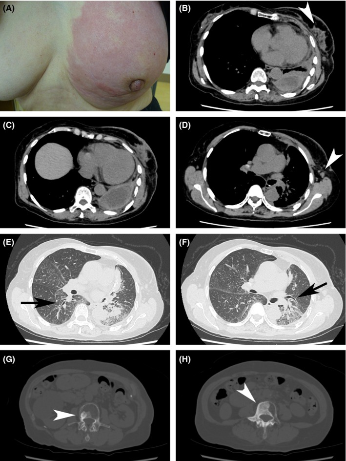
A, Left breast showing diffuse erythema swelling. B‐D, CT image obtained with mediastinal window settings. B, Irregular mass and satellite nodules (white arrowhead). B and C, Pleural effusion/thickening. D, Axillary lymph nodes (white arrowhead). E and F, CT images obtained with mediastinal window settings showing thickened bronchovascular bundles, indicating lung cancer aggravation (arrows). G and H, CT images obtained with bone settings showing multiple bone metastases (white arrowheads)
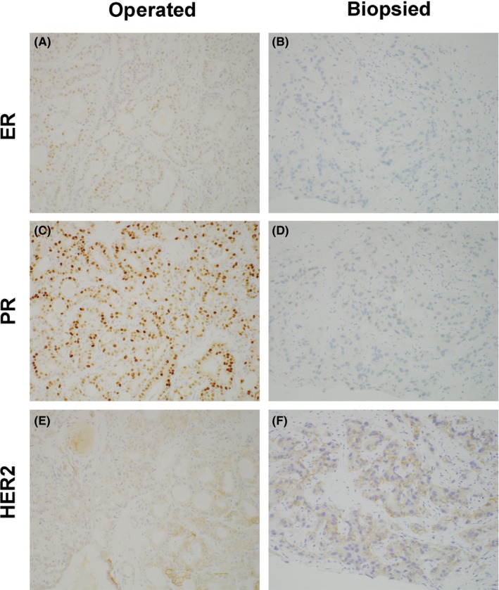
Expression of ER, PR, and HER2 in the breast cancer tissue from surgery and from core needle biopsy. (A, C, E) Breast cancer tissue from surgery (×200); (B, D, F) biopsied breast cancer tissue (×200). (A, B) ER, (B, E) PR, and (C, F) HER2. In the breast cancer tissue from surgery, ER was barely positive, PR was positive, and HER2 was 1+. In the breast cancer tissue from core needle biopsy, ER and PR were negative, and HER2 was 1+
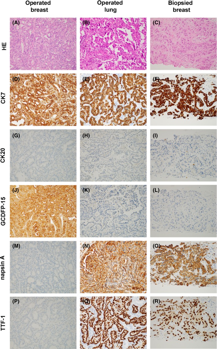
IHC analyses of the breast cancer tissue from surgery, lung cancer tissue from surgery, and breast cancer tissue from core needle biopsy. (A, D, G, J, M, P) Breast cancer tissue from surgery (×200); (B, E, H, K, N, Q); lung cancer tissue from surgery (×200); (C, F, I, L, O, R) breast cancer tissue from the core needle biopsy (×200). (A, B, C) HE, (D, E, G) CK7, (G, H, I) CK20, (J, K, L) GCDFP‐15, (M, N, O) napsin A, and (P, Q, R) TTF‐1. HE; hematoxylin and eosin. As shown in figure, the IHC staining pattern of the breast cancer tissue from core needle biopsy was the same that of the lung cancer tissue from surgery but was not the same as that of the breast cancer tissue from surgery
References
-
- Lee SK, Kim WW, Kim SH, et al. Characteristics of metastasis in the breast from extramammary malignancies. J Surg Oncol. 2010;101:137‐140. - PubMed
-
- Vizcaíno I, Torregrosa A, Higueras V, et al. Metastasis to the breast from extramammary malignancies: a report of four cases and a review of literature. Eur Radiol. 2001;11:1659‐1665. - PubMed
-
- Jochimsen PR, Brown RC. Metastatic melanoma in the breast masquerading as fibroadenoma. JAMA. 1976;236:2779‐2780. - PubMed
-
- Lee SH, Park JM, Kook SH, Han BK, Moon WK. Metastatic tumors to the breast: mammographic and ultrasonographic findings. J Ultrasound Med. 2000;19:257‐262. - PubMed
Publication types
LinkOut - more resources
Full Text Sources
Other Literature Sources
Research Materials
Miscellaneous
