

Spontaneous Lung Herniation Leading to Extensive Subcutaneous Emphysema, Pneumothorax, Pneumomediastinum, and Pneumopericardium
- PMID: 30148014
- PMCID: PMC6107042
- DOI: 10.7759/cureus.2861
Spontaneous Lung Herniation Leading to Extensive Subcutaneous Emphysema, Pneumothorax, Pneumomediastinum, and Pneumopericardium
Abstract
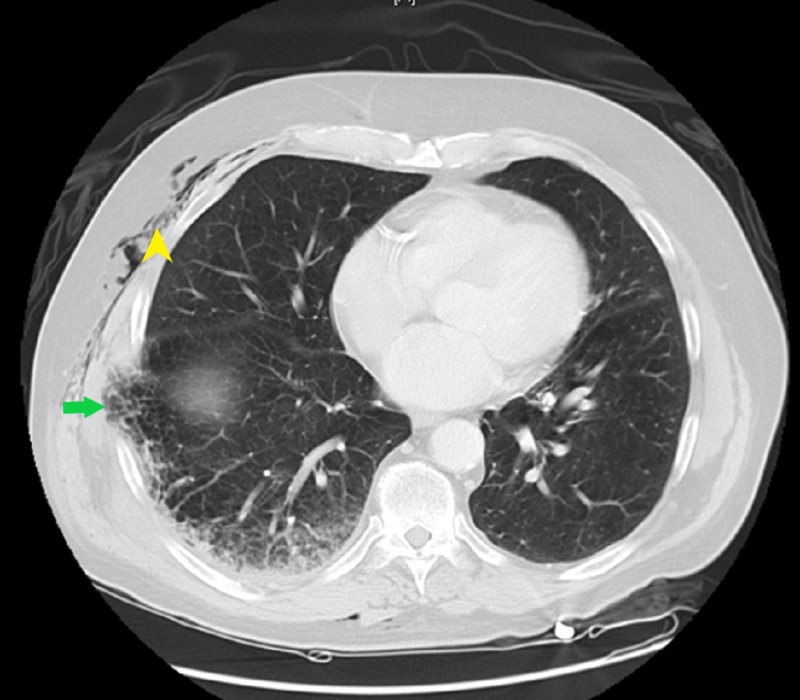
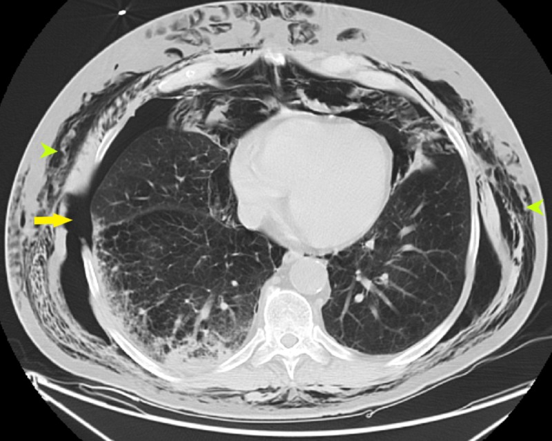
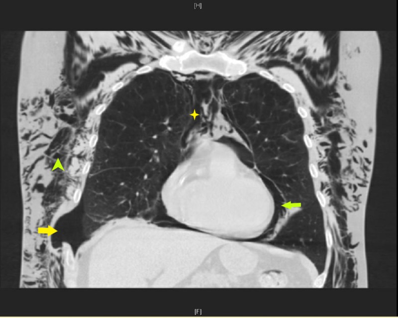
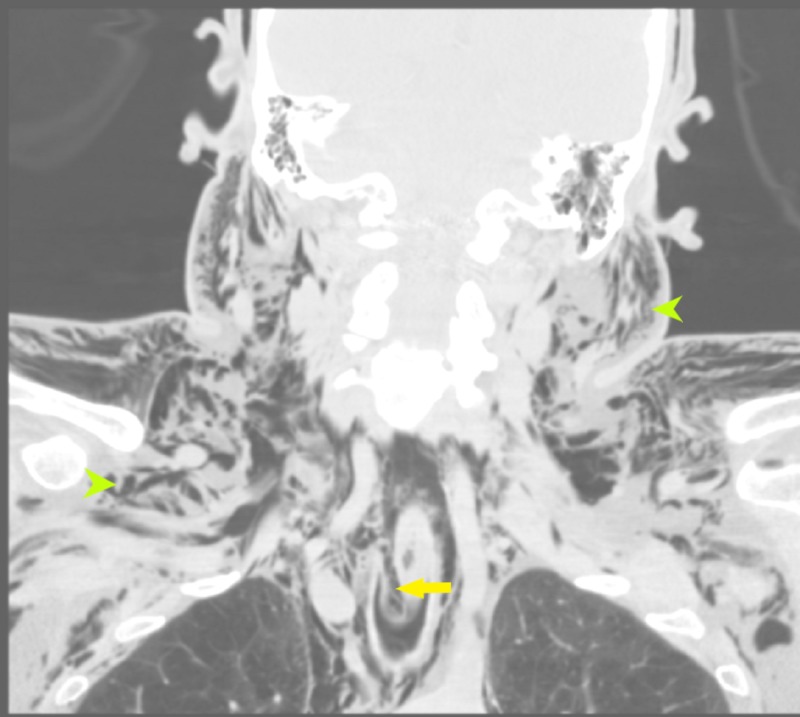
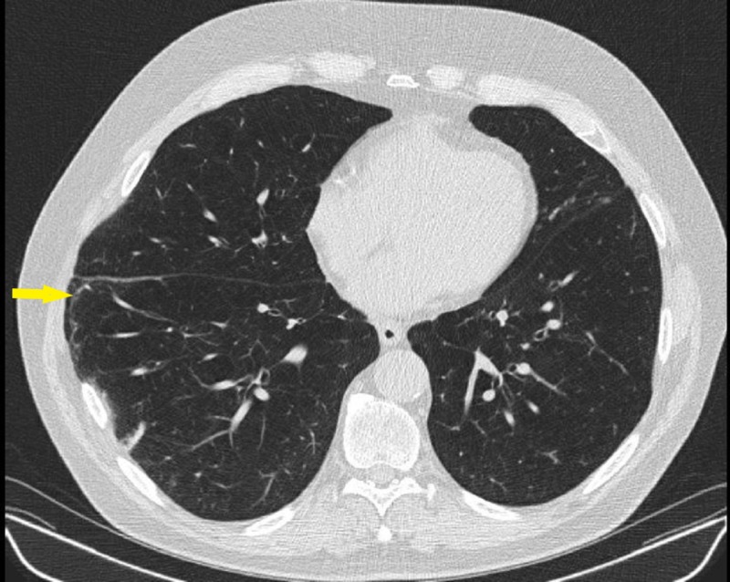
Spontaneous lung herniation is a rare phenomenon in which the lung parenchyma along with the pleural membranes protrudes outside their usual boundaries and can lead to a wide variety of complications. We are reporting a case of a middle-aged male who presented with chronic obstructive pulmonary disease (COPD) exacerbation with severe bouts of cough. Initial computed tomography (CT) chest was unrevealing, but two days later, he developed spontaneous lung herniation, which was initially managed conservatively, but later it progressed to pneumothorax, pneumomediastinum, with striking CT scan images showing extensive subcutaneous emphysema. Blowhole incisions were done on the anterior chest wall which led to ultimate recovery.
Keywords: blow-hole incisions; diaphragmatic hernia.
Conflict of interest statement
The authors have declared that no competing interests exist.
Figures
References
-
- Tack D, Wattiez A, Schtickzelle JC, Delcour C. Eur Radiol. Vol. 10. Accessed Jun; 2000. Spontaneous lung herniation after a single cough; pp. 500–502. - PubMed
-
- Cough-induced intercostal lung herniation requiring surgery: report of a case. Sulaiman A, Cottin V, De Souza Neto EP, Orsini A, Cordier JF, Gamondes JP, Tronc F. Surgery. 2006;36:978–980. - PubMed
-
- A painful sneeze: spontaneous thoracic lung herniation induced by vigorous sneeze. Bhardwaj H, Bhardwaj B, Youness HA. J Bronchology Interv Pulmonol. 2014;21:61–64. - PubMed
-
- Spontaneous lung herniation through the chest wall. Cox M, Thota D, Trevino R. Mil Med Res. 2018;183:233–234. - PubMed
-
- Intercostal lung hernia: radiographic and MDCT findings. Zia Z, Bashir O, Ramjas GE, Kumaran M, Pollock JG, Pointon K. Clin Radiol. 2013;68:412–417. - PubMed
Publication types
LinkOut - more resources
Full Text Sources
Other Literature Sources