. 2018 Aug 17;4(3):193-196.
doi: 10.1016/j.jvscit.2018.03.010. eCollection 2018 Sep.

Chylothorax secondary to venous outflow obstruction treated with transcervical retrograde thoracic duct cannulation with embolization and venous reconstruction

Affiliations

Chylothorax secondary to venous outflow obstruction treated with transcervical retrograde thoracic duct cannulation with embolization and venous reconstruction

Jeffrey Forris Beecham Chick et al. J Vasc Surg Cases Innov Tech. .

Abstract

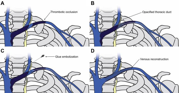
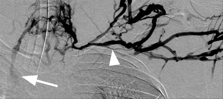
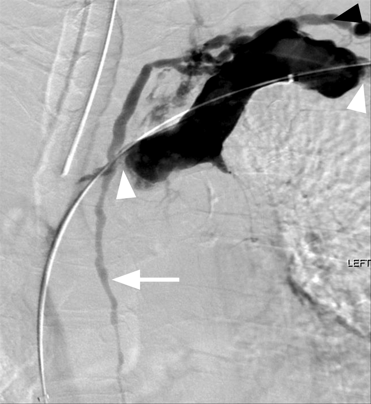
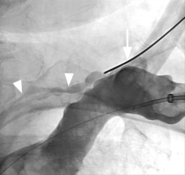
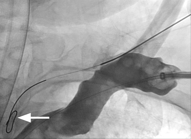
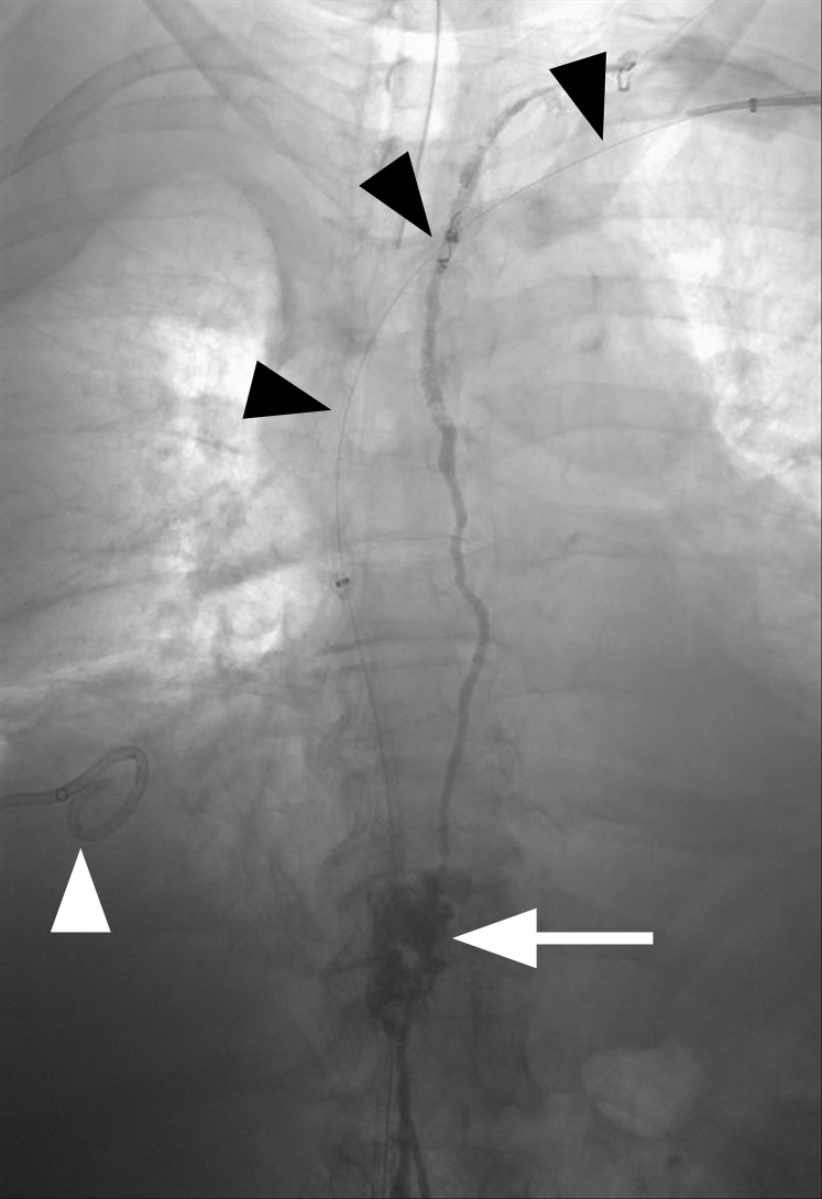
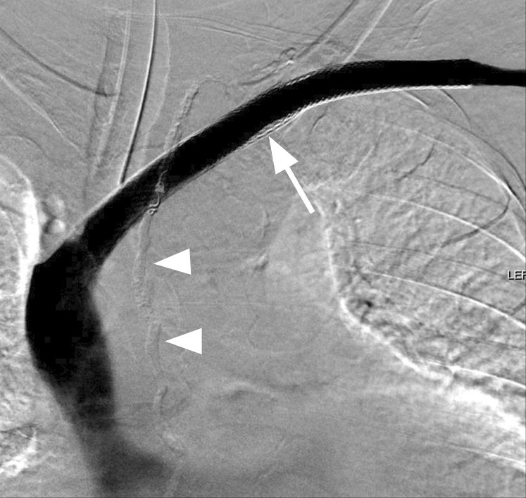
A chylothorax may be due to either direct trauma or occlusion of the thoracic duct. Treatments include antegrade or retrograde glue and coil embolization as well as thoracic duct stent graft placement. This report describes a patient with chylothorax secondary to venous outflow occlusion. Left upper extremity venography demonstrated multifocal left brachiocephalic and axillary vein occlusions with retrograde filling of an engorged and disrupted thoracic duct. Retrograde thoracic duct lymphangiography with embolization and left upper extremity venous reconstruction were performed with complete resolution of chylothorax.

Keywords: Central venous occlusion; Chylothorax; Embolization; Retrograde; Thoracic duct; Venous reconstruction.


Figures

Fig 1
Fig 1
Schematic illustration of transcervical retrograde thoracic duct glue embolization and venous reconstruction. A, Thrombotic occlusion of the left axillary and brachiocephalic veins. B, Opacification of the proximal thoracic duct through pressurized venography. C, Transcervical access and glue embolization of the thoracic duct. D, Venous recanalization and stenting of the left axillary and distal brachiocephalic veins.
Fig 2
Fig 2
Frontal digital subtraction venography of the left arm showing complete occlusion of the left axillary and brachiocephalic veins (arrowhead). Numerous collateral veins are seen in the upper chest that reconstitute the superior vena cava (arrow).
Fig 3
Fig 3
After successful crossing of the left axillary and brachiocephalic vein occlusions, pressurized digital subtraction venography within the excluded subclavian vein shows retrograde filling of the thoracic duct (arrow). Tandem occlusions are noted in the axillary and brachiocephalic veins (white arrowheads). The left internal jugular vein is occluded. Contrast material is seen refluxing into a straight-appearing segment of a left lymphatic duct (black arrowhead) and the main thoracic duct.
Fig 4
Fig 4
After multiple unsuccessful transvenous attempts to cannulate the terminal thoracic duct (arrowheads) with various reverse curve catheters, a 21-gauge Chiba needle (arrow) was used to access a lymphatic channel under fluoroscopic guidance.
Fig 5
Fig 5
Once access was obtained, an 0.018-inch Nitrex wire was navigated into the main thoracic duct (arrow).
Fig 6
Fig 6
Frontal fluoroscopic image showing n-butyl cyanoacrylate filling the thoracic duct. The area of disruption within the thoracic duct, likely from central venous occlusion, is seen with glue filling this segment (arrow). A wire is identified crossing the central venous occlusions (black arrowheads). A pigtail catheter is visualized in the right pleural space (white arrowhead).
Fig 7
Fig 7
Frontal left upper extremity venogram showing successful placement of Wallstents in the brachiocephalic and subclavian veins (arrow) with free flow of contrast material from the left arm to the superior vena cava. Stents were extended into the left axillary vein to ensure adequate inflow. Glue is seen within the thoracic duct (arrowheads).

References

    1. Maldonado F., Cartin-Ceba R., Hawkins F.J., Ryu J.H. Medical and surgical management of chylothorax and associated outcomes. Am J Med Sci. 2010;339:314–318. - PubMed
    1. Chen E., Itkin M. Thoracic duct embolization for chylous leaks. Semin Intervent Radiol. 2011;28:63–74. - PMC - PubMed
    1. Pamarthi V., Stecker M.S., Schenker M.P., Baum R.A., Killoran T.P., Suzuki Han A. Thoracic duct embolization and disruption for treatment of chylous effusions: experience with 105 patients. J Vasc Interv Radiol. 2014;25:1398–1404. - PubMed
    1. Johnson O.W., Chick J.F., Chauhan N.R., Fairchild A.H., Fan C.M., Stecker M.S. The thoracic duct: clinical importance, anatomic variation, imaging, and embolization. Eur Radiol. 2016;26:2482–2493. - PubMed
    1. Doerr C.H., Allen M.S., Nichols F.C., Ryu J.H. Etiology of chylothorax in 203 patients. Mayo Clin Proc. 2005;80:867–870. - PubMed

LinkOut - more resources