. 2018 Jun:2:3.
doi: 10.21037/mps.2018.05.01. Epub 2018 Jun 11.

Integrating Mass Spectrometry with Microphysiological Systems for Improved Neurochemical Studies

Affiliations

Integrating Mass Spectrometry with Microphysiological Systems for Improved Neurochemical Studies

Emily G Tillmaand et al. Microphysiol Syst. 2018 Jun.

Abstract

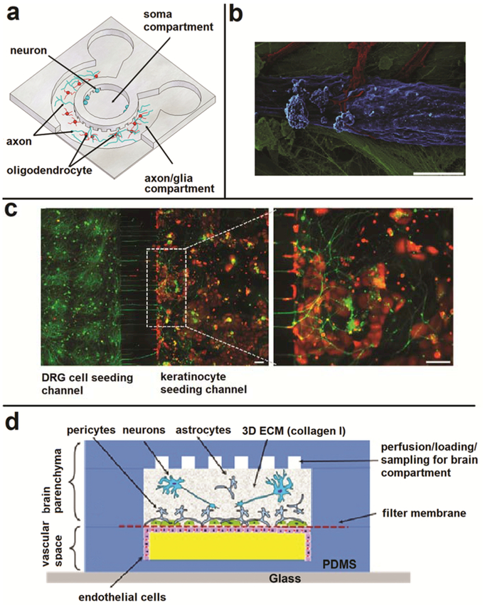
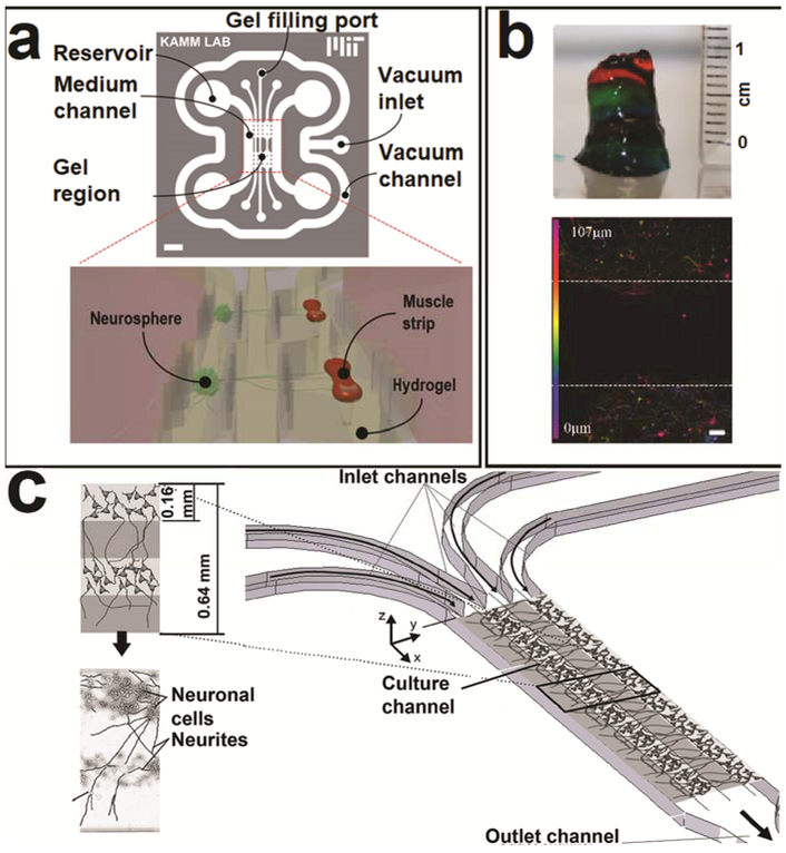
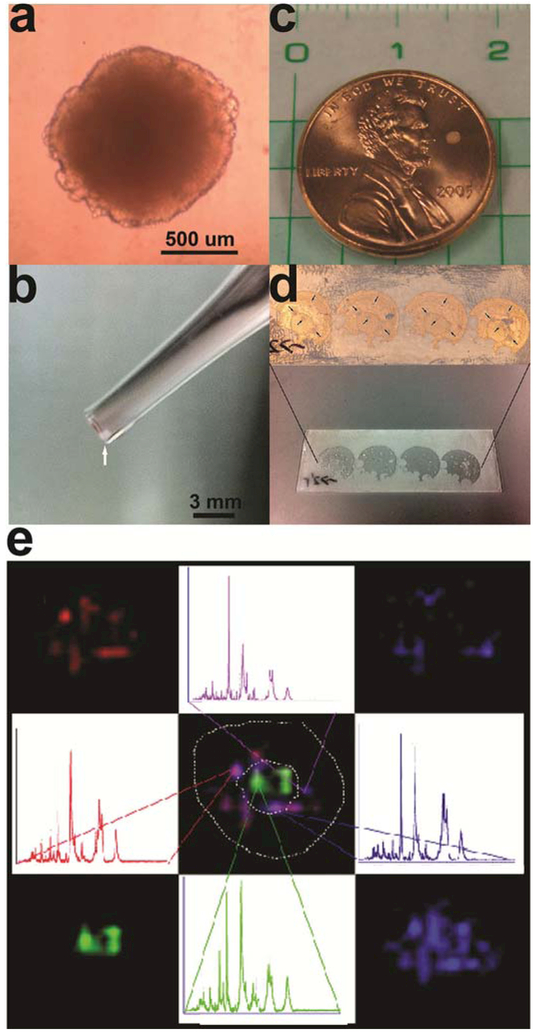
Microphysiological systems, often referred to as "organs-on-chips", are in vitro platforms designed to model the spatial, chemical, structural, and physiological elements of in vivo cellular environments. They enhance the evaluation of complex engineered biological systems and are a step between traditional cell culture and in vivo experimentation. As neurochemists and measurement scientists studying the molecules involved in intercellular communication in the nervous system, we focus here on recent advances in neuroscience using microneurological systems and their potential to interface with mass spectrometry. We discuss a number of examples - microfluidic devices, spheroid cultures, hydrogels, scaffolds, and fibers - highlighting those that would benefit from mass spectrometric technologies to obtain improved chemical information.


Conflict of interest statement

Conflict of Interest The authors declare no conflict of interest.

Figures

Figure 1:
Figure 1:. Microfluidic devices for the creation of microneurological systems.
(a) Schematic illustration of the microfluidic compartmentalized CNS neuron co-culture platform. A 3D view of the circular device. Reproduced from Park J, Koito H, Li J, et al. Microfluidic Compartmentalized Co-Culture Platform for CNS Axon Myelination Research. Biomed Microdevices 2009;11:1145. Copyright (2009) with permission of Springer (59). (b) False-colored SEM image showing motor neurons (red) interacting with myotubules (blue) in the distal chamber of a microfluidic device. Scale bar: 5 μm. Reprinted from Southam KA, King AE, Blizzard CA, et al. Microfluidic Primary Culture Model of the Lower Motor Neuron–Neuromuscular Junction Circuit. J Neurosci Methods 2013;218:164-9. Copyright (2013) with permission from Elsevier (61). (c) Co-culture of rat neonatal keratinocytes (stained for cytokeratin 5, red) with rat neonatal primary sensory neurons (stained for B3 tubulin, green) in a dual chamber device. Scale bar: 100 μm. Adapted from Tsantoulas C, Farmer C, Machado P, et al. Probing Functional Properties of Nociceptive Axons Using a Microfluidic Culture System. PLoS One 2013;8:e80722. Provided under Creative Commons license 3.0 (71). (d) Schematic of a neurovascular unit created within a microfluidic device. Adapted from Brown JA, Pensabene V, Markov DA, et al. Recreating Blood-Brain Barrier Physiology and Structure on Chip: A Novel Neurovascular Microfluidic Bioreactor. Biomicrofluidics 2015;9:054124. Provided under Creative Commons license 3.0. (79).
Figure 2:
Figure 2:. The use of neurospheres and hydrogels in microphysiological model systems.
(a) Schematic of the integration of neurosphere culture within a microfluidic device. Adapted from Uzel SGM, Platt RJ, Subramanian V, et al. Microfluidic Device for the Formation of Optically Excitable, Three-Dimensional, Compartmentalized Motor Units. Science Advances 2016;2. Provided under Creative Commons Attribution-Noncommercial 4.0 International license. http://advances.sciencemag.Org/content/2/8/e1501429.full (65). (b) Hydrogel layered cortex models. Top: 3D printed hydrogel layers Bottom: fluorescent images showing the layers with and without cells. The scale bar represents 100 μm. Reprinted from Lozano R, Stevens L, Thompson BC, et al. 3D Printing of Layered Brain-Like Structures Using Peptide Modified Gellan Gum Substrates. Biomaterials 2015;67:264-73, Copyright (2015) with permission from Elsevier (87). (c) Schematic of a microfluidic device used to create hydrogel layers imitating the cortex. Reprinted from Kunze A, Giugliano M, Valero A, et al. Micropatterning Neural Cell Cultures in 3D with a Multi-Layered Scaffold. Biomaterials 2011;32:2088-98. Copyright (2011) with permission from Elsevier (88).
Figure 3:
Figure 3:. MS imaging of spherical cultures.
(a) A spherical liver cell culture. (b) Arrow is pointing to the spherical culture in a pipet tip. (c) Spherical culture size compared to a penny (the white spot next to Lincoln’s nose). (d) Spherical cultures imbedded in gelatin, sectioned, and placed on an indium tin oxide-coated microscope slide. The areas consisting of the cell culture slices are small and are located at the tips of the small black lines in the top image. (e) Representative mass spectra and corresponding ion intensity maps for MSI on the spherical culture. Adapted with permission from Li H, Hummon AB. Imaging Mass Spectrometry of Three-Dimensional Cell Culture Systems. Anal Chem 2011;83:8794-801, ref (54). Copyright 2011 American Chemical Society.

References

    1. Wikswo JP. The Relevance and Potential Roles of Microphysiological Systems in Biology and Medicine. Exp Biol Med (Maywood) 2014;239:1061–72. - PMC - PubMed
    1. Baker BM, Chen CS. Deconstructing the Third Dimension – How 3D Culture Microenvironments Alter Cellular Cues. J Cell Sci 2012;125:3015–24. - PMC - PubMed
    1. Huang S, Wikswo J. Dimensions of Systems Biology Reviews of Physiology Biochemistry and Pharmacology. Berlin, Heidelberg: Springer; 2007. p. 81–104. - PubMed
    1. Li X, Valadez AV, Zuo P, et al. Microfluidic 3D Cell Culture: Potential Application for Tissue-Based Bioassays. Bioanalysis 2012;4:1509–25. - PMC - PubMed
    1. Baharvand H, Hashemi SM, Kazemi Ashtiani S, et al. Differentiation of Human Embryonic Stem Cells into Hepatocytes in 2D and 3D Culture Systems in Vitro. Int J Dev Biol 2006;50:645–52. - PubMed

LinkOut - more resources