

Effect of total sonicated Aggregatibacter actinomycetemcomitans fragments on gingival stem/progenitor cells
- PMID: 30148477
- PMCID: PMC6167108
- DOI: 10.4317/medoral.22661
Effect of total sonicated Aggregatibacter actinomycetemcomitans fragments on gingival stem/progenitor cells
Abstract
Background: Aggregatibacter-actinomycetemcomitans (A.actinomycetemcomitans) are strongly associated with localized-aggressive-periodontitis (LAgP). The study's aim was to test for the first time the effect of total sonicated A.actinomycetemcomitans-bacterial-fragments on gingival mesenchymal stem/progenitor cells' (G-MSCs) proliferation and regenerative gene expression in-vitro.
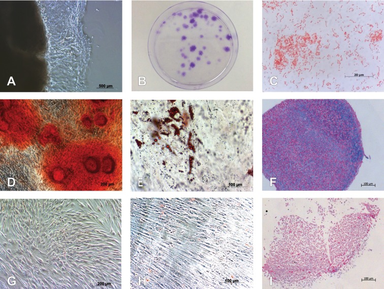
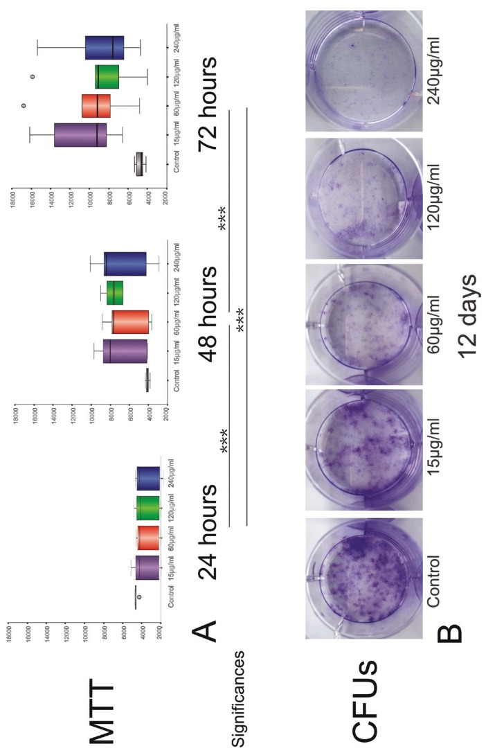
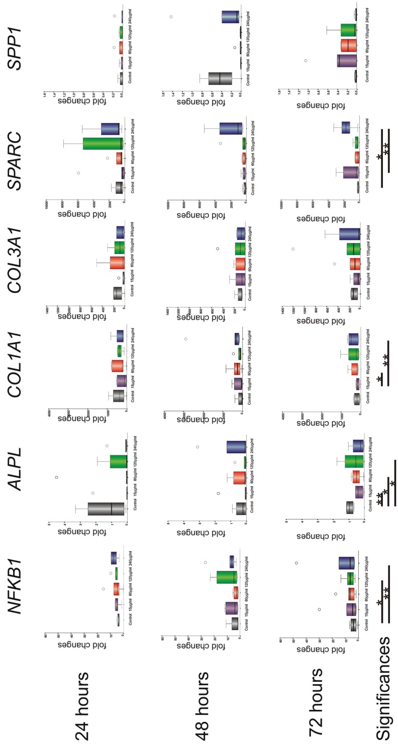
Material and methods: G-MSCs were isolated, characterized, expanded and stimulated by total sonicated A.actinomycetemcomitans-bacterial-fragments (0 (negative-control), 15, 60, 120 and 240µg/ml; serovar-b; n=6/group). Cellular proliferation and NF-κβ (NFKB1), Alkaline Phosphatase (ALPL), Collagen-I (COL1A1), Collagen-III (COL3A1), Osteonectin (SPARC) and Osteopontin (SPP1) m-RNA expression were assessed via reverse-transcription-polymerase-chain-reaction (RT-PCR) at 24, 48 and 72 hours and CFUs-ability evaluated at twelve days.
Results: G-MSCs demonstrated stem/progenitor cells' characteristics. A.actinomycetemcomitans-bacterial-fragments (up to 72 hours) resulted in marked G-MSCs' proliferation over-time (p<0.001) and elevated NFKB1 (p=0.017), COL1A1 (p=0.025), SPARC (p=0.025), decreased ALPL (p=0.017), with no significant differences for COL3A1 and SPP1 expression or stimulation times (p>0.05; Friedman-test). Longer-term stimulation for twelve days reduced G-MSCs' CFUs.
Conclusions: Sonicated A.actinomycetemcomitans-bacterial-fragments' exert beneficial short-term effects on G-MSCs' proliferative and non-mineralized tissue forming aptitude. Results shed new light on the importance of periodontal treatment for LAgP patients, using power driven sonic/ultrasonic devices, which, in addition to reducing the subgingival microbial load, produces cell-stimulatory A.actinomycetemcomitans-bacterial-fragments, with positive attributes on tissue reparative/regenerative responses of tissue resident stem/progenitor cells in their niche.
Conflict of interest statement
Figures
References
-
- Page RC, Offenbacher S, Schroeder HE, Seymour GJ, Kornman KS. Advances in the pathogenesis of periodontitis: summary of developments, clinical implications and future directions. Periodontol 2000. 1997;14:216–48. - PubMed
-
- Armitage GC. Development of a classification system for periodontal diseasesand conditions. Northwest Dent. 2000;79:31–5. - PubMed
-
- Albandar JM, Tinoco EMB. Global epidemiology of periodontal diseases in children and young persons. Periodontol 2000. 2002;29:153–76. - PubMed
-
- Diehl SR, Wu T, Michalowicz BS, Brooks CN, Califano JV, Burmeister JA. Quantitative measures of aggressive periodontitis show substantial heritability and consistency with traditional diagnoses. J Periodontol. 2005;76:279–88. - PubMed
-
- Albandar JM. Aggressive and acute periodontal diseases. Periodontol 2000. 2014;65:7–12. - PubMed
MeSH terms
Substances
LinkOut - more resources
Full Text Sources
Other Literature Sources
Medical
Research Materials
Miscellaneous
