. 2018 Aug 27;13(8):e0201948.
doi: 10.1371/journal.pone.0201948. eCollection 2018.

Chemoproteomic identification of molecular targets of antifungal prototypes, thiosemicarbazide and a camphene derivative of thiosemicarbazide, in Paracoccidioides brasiliensis

Affiliations

Chemoproteomic identification of molecular targets of antifungal prototypes, thiosemicarbazide and a camphene derivative of thiosemicarbazide, in Paracoccidioides brasiliensis

Joyce Villa Verde Bastos Borba et al. PLoS One. .

Abstract

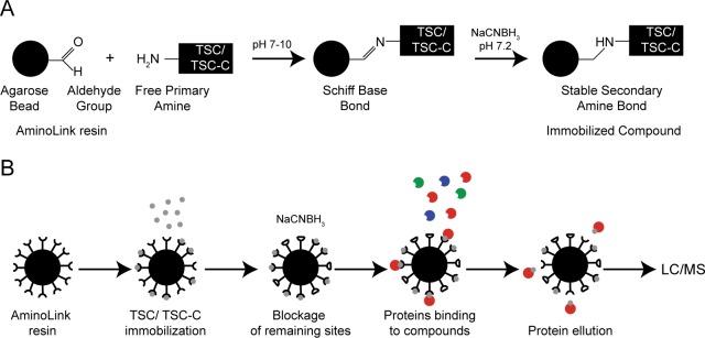
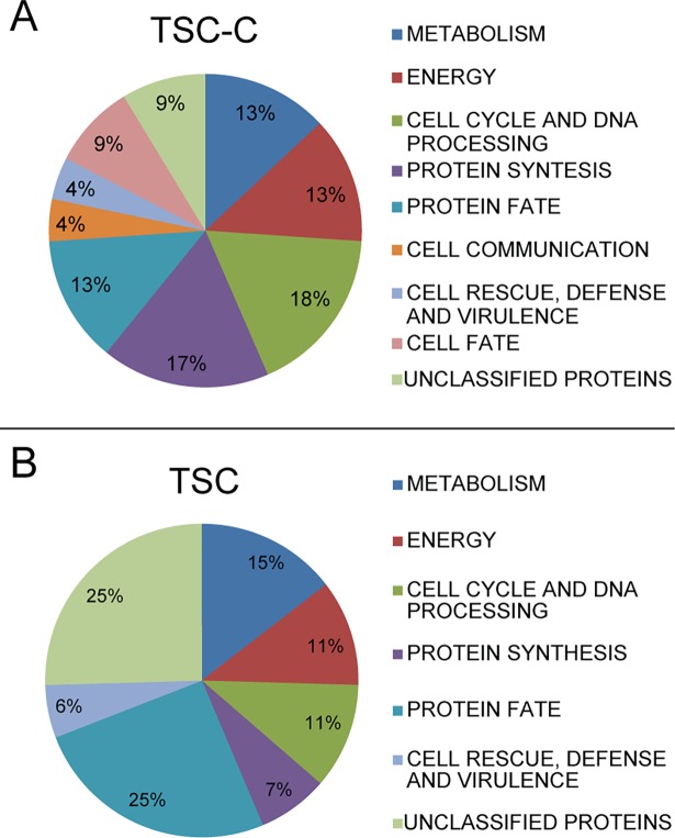
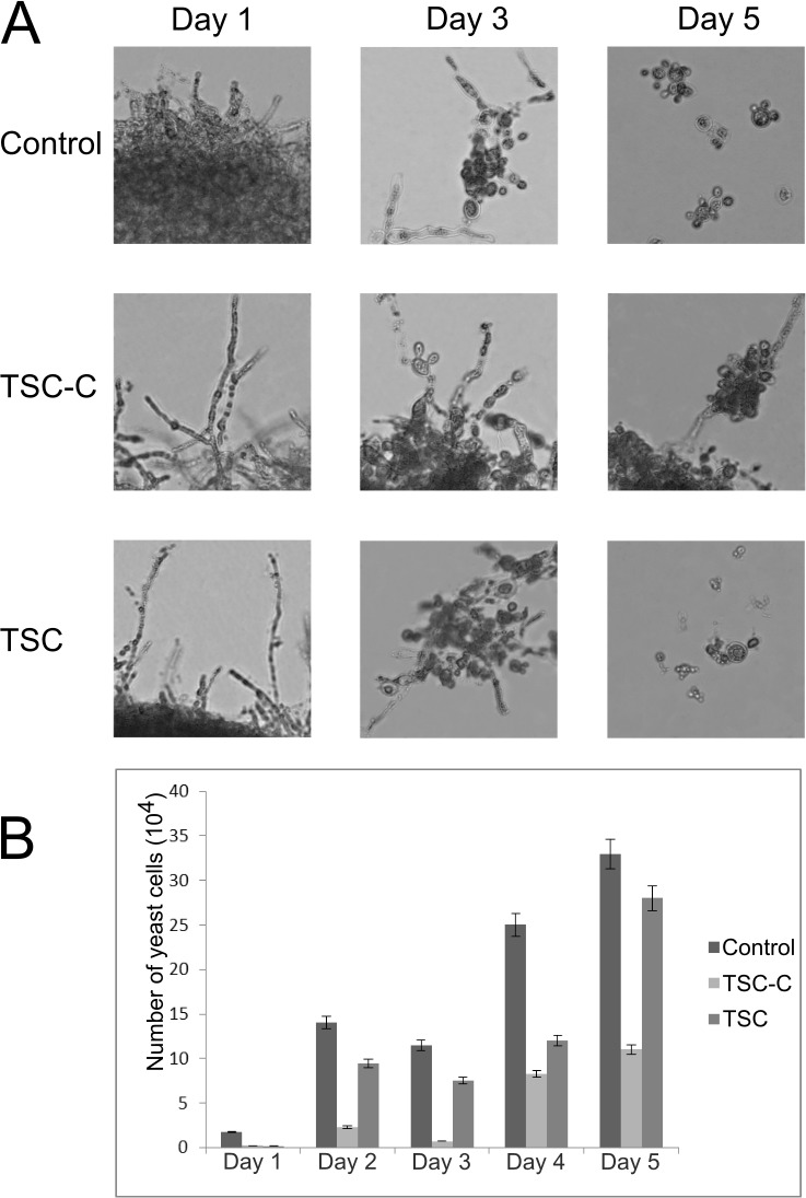
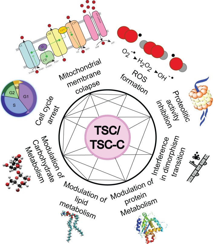
Paracoccidioidomycosis (PCM) is a neglected human systemic disease caused by species of the genus Paracoccidioides. The disease attacks the host's lungs and may disseminate to many other organs. Treatment involves amphotericin B, sulfadiazine, trimethoprim-sulfamethoxazole, itraconazole, ketoconazole, or fluconazole. The treatment duration is usually long, from 6 months to 2 years, and many adverse effects may occur in relation to the treatment; co-morbidities and poor treatment adherence have been noted. Therefore, the discovery of more effective and less toxic drugs is needed. Thiosemicarbazide (TSC) and a camphene derivative of thiosemicarbazide (TSC-C) were able to inhibit P. brasiliensis growth at a low dosage and were not toxic to fibroblast cells. In order to investigate the mode of action of those compounds, we used a chemoproteomic approach to determine which fungal proteins were bound to each of these compounds. The compounds were able to inhibit the activities of the enzyme formamidase and interfered in P. brasiliensis dimorphism. In comparison with the transcriptomic and proteomic data previously obtained by our group, we determined that TSC and TSC-C were multitarget compounds that exerted effects on the electron-transport chain and cell cycle regulation, increased ROS formation, inhibited proteasomes and peptidases, modulated glycolysis, lipid, protein and carbohydrate metabolisms, and caused suppressed the mycelium to yeast transition.


Conflict of interest statement

The authors have declared that no competing interests exist.

Figures

Fig 1
Fig 1. AminoLink resin protocol.
Molecular view of the immobilization step (A) and overall view of the role protocol (B) AminoLink resins contain aldehyde groups that reacted with the free amino groups of TSC or TSC-C. The aldehyde sites that do not react with the compound are blocked with sodium cyanoborohydride (A). After compound immobilization, the fungal protein extract is added to the resin column. The proteins that interact with the compounds stay in the resin and the rest of the proteins are washed away. The interacting proteins are eluted from the resin, digested, and identified through mass spectrometry (B).
Fig 2
Fig 2. The graph indicates the statistically enriched MIPS functions.
Proteins that were bound to TSC-C and TSC. The functional classification was based on the MIPS functional annotation scheme. Each functional class is represented as a color-coded segment and expressed as a percentage of the total number of proteins.
Fig 3
Fig 3. Effects of TSC and TSC-C on P. brasiliensis transition from mycelium to yeast.
Mycelium cells were incubated for 5 days at 36°C on medium containing 85.3 μM TSC or 17.2 μM TSC-C. Cell morphology was observed by optical microscopy (A). Yeast cells were counted by using a Neubauer chamber (B).
Fig 4
Fig 4. Main cellular processes influenced by TSC and TSC-C based on a comparison between the transcriptomic, proteomic, and chemoproteomic data.

References

    1. Turissini DA, Gomez OM, Teixeira MM, McEwen JG, Matute DR. Species boundaries in the human pathogen Paracoccidioides. Fungal Genet Biol. Elsevier; 2017;106: 9–25. 10.1016/j.fgb.2017.05.007 - DOI - PMC - PubMed
    1. Desjardins C a, Champion MD, Holder JW, Muszewska A, Goldberg J, Bailão AM, et al. Comparative genomic analysis of human fungal pathogens causing paracoccidioidomycosis. PLoS Genet. 2011;7: e1002345 10.1371/journal.pgen.1002345 - DOI - PMC - PubMed
    1. Teixeira MM, Theodoro RC, de Carvalho MJA, Fernandes L, Paes HC, Hahn RC, et al. Phylogenetic analysis reveals a high level of speciation in the Paracoccidioides genus. Mol Phylogenet Evol. 2009;52: 273–283. 10.1016/j.ympev.2009.04.005 - DOI - PubMed
    1. Bocca AL, Amaral AC, Teixeira MM, Sato PK, Shikanai-Yasuda MA, Soares Felipe MS. Paracoccidioidomycosis: eco-epidemiology, taxonomy and clinical and therapeutic issues. Future Microbiol. 2013;8: 1177–1191. 10.2217/fmb.13.68 - DOI - PubMed
    1. Shikanai-Yasuda MA, Mendes RP, Colombo AL, Queiroz-Telles F de, Kono ASG, Paniago AMM, et al. Brazilian guidelines for the clinical management of paracoccidioidomycosis. Rev Soc Bras Med Trop. 2017;50: 715–740. 10.1590/0037-8682-0230-2017 - DOI - PubMed

Publication types

MeSH terms