Observational Study
. 2018 Aug 27;13(8):e0202950.
doi: 10.1371/journal.pone.0202950. eCollection 2018.

Progression of cardiac allograft vasculopathy assessed by serial three-vessel quantitative coronary angiography

Affiliations
Observational Study

Progression of cardiac allograft vasculopathy assessed by serial three-vessel quantitative coronary angiography

Christian Zanchin et al. PLoS One. .

Abstract

Background: The purpose of the present study was to assess the short- and long-term progression of cardiac allograft vasculopathy (CAV) using serial 3-vessel quantitative coronary angiography (QCA).

Methods: CAV progression was assessed using serial 3-vessel QCA analysis at baseline, 1-year and long-term angiographic follow-up (8.5±3.7 years) after heart transplantation. The change in minimal lumen diameter (MLD) and percent diameter stenosis (%DS) was serially assessed within matched segments. Patients were graded according to the ISHLT-CAV classification and grouped as ISHLT-CAV0 and ISHLT-CAV1-3. The primary endpoint was mean change in MLD and %DS.

Results: A total of 41 patients and 520 matched segments were available for serial 3-vessel QCA. Overall, MLD decreased non-significantly from baseline to 1-year follow-up and significantly from 1-year to the long-term angiographic follow-up (Δ-0.08mm/year [95%CI -0.11 to -0.05], P<0.001). %DS increased significantly from baseline to 1-year (Δ+0.96%/year [95%CI 0.04 to 1.88], P = 0.041) and from 1-year to long-term angiographic follow-up (Δ+0.61%/year [95%CI 0.33 to 0.88], P<0.001). ISHLT-CAV1-3 at 1 year and at long-term angiographic follow-up was observed in 22% and 61%, respectively. Between baseline and long-term angiographic follow-up, a significant reduction in MLD was observed within both groups without a significant difference in the reduction between the two groups (ISHLT-CAV0: median -0.49mm [IQR -0.54 to -0.43] vs. ISHLT-CAV1-3: median -0.40mm [IQR -0.44 to -0.35], P = 0.4).

Conclusion: The current data suggest that QCA can't predict CAV beyond 1 year, but, QCA affirmed that CAV progresses to a similar extent in patients who do not develop visual CAV during long-term follow-up.


Conflict of interest statement

The authors have declared that no competing interests exist.

Figures
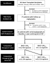
Fig 1
Fig 1. Flow chart.
CAV indicates cardiac allograft vasculopathy; FUP, follow-up; QCA, quantitative coronary angiography; ISHLT, International Society of Heart and Lung Transplantation.
Fig 2
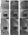
Fig 2. Serial 3-vessel quantitative coronary angiography analysis.
This figure shows the serial quantitative coronary angiography analysis within matched regions of all coronary artery segments at baseline, at 1 year follow-up, and at latest available angiographic follow-up. Coronary artery segments were classified according to the modified AHA/ACC classification. MLD indicates minimal lumen diameter; %DS, percent diameter stenosis.
Fig 3
Fig 3. Serial 3-vessel quantitative coronary angiography analysis.
Box-plot representation of minimal lumen diameter and maximal percent diameter stenosis at baseline (median 0.14 years [0.12 to 0.16], at 1 year (median 1.17 years [1.14 to 1.20]) and at long-term angiographic follow-up (median 8.61 years [8.24 to 8.99]) after HTx. Lower and upper box edges are the quartiles and thick line is the median.
Fig 4
Fig 4. Quantitative angiographic analysis.
Box-plot representation of the per-patient mean angiographic change in minimal lumen diameter (minimal lumen diameter 1 year—minimal lumen diameter baseline and minimal lumen diameter latest angiographic follow-up—minimal lumen diameter baseline) from coronary artery segments that were serially assessed and matched. The analysis is stratified according to absence (n = 16) or presence (n = 25) of ISHLT-CAV at 1 year, and at latest angiographic follow-up (median 8.61 years [8.24 to 8.99]). Lower and upper box edges are the quartiles and thick line is the median. A horizontal reference line at change = 0 is drawn.

Similar articles

Cited by

References

    1. Lund LH, Edwards LB, Kucheryavaya AY, Benden C, Dipchand AI, Goldfarb S, et al. The Registry of the International Society for Heart and Lung Transplantation: Thirty-second Official Adult Heart Transplantation Report—2015; Focus Theme: Early Graft Failure. J Hear Lung Transplant. Elsevier; 2015;34: 1244–1254. 10.1016/j.healun.2015.08.003 - DOI - PubMed
    1. Rahmani M, Cruz RP, Granville DJ, McManus BM. Allograft vasculopathy versus atherosclerosis. Circ Res. 2006;99: 801–15. 10.1161/01.RES.0000246086.93555.f3 - DOI - PubMed
    1. Clerkin KJ, Ali ZA, Mancini DM. New developments for the detection and treatment of cardiac vasculopathy. Curr Opin Cardiol. 2017;32: 316–325. 10.1097/HCO.0000000000000388 - DOI - PMC - PubMed
    1. Ruygrok PN, Webber B, Faddy S, Muller DW, Keogh A. Angiographic regression of cardiac allograft vasculopathy after introducing sirolimus immunosuppression. J Heart Lung Transplant. 2003;22: 1276–9. 10.1016/S1053-2498(02)01239-1 - DOI - PubMed
    1. Costanzo MR, Dipchand A, Starling R, Anderson A, Chan M, Desai S, et al. The International Society of Heart and Lung Transplantation Guidelines for the care of heart transplant recipients. J Heart Lung Transplant. Elsevier Inc.; 2010;29: 914–56. 10.1016/j.healun.2010.05.034 - DOI - PubMed

Publication types

MeSH terms