Review
. 2018 Oct:72:18-41.
doi: 10.1016/j.plipres.2018.08.002. Epub 2018 Aug 25.

Properties, metabolism and roles of sulfogalactosylglycerolipid in male reproduction

Affiliations
Review

Properties, metabolism and roles of sulfogalactosylglycerolipid in male reproduction

Nongnuj Tanphaichitr et al. Prog Lipid Res. 2018 Oct.

Abstract

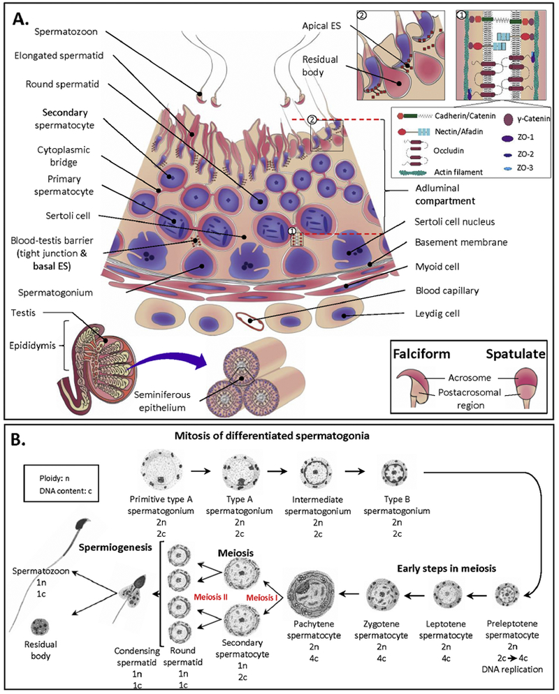
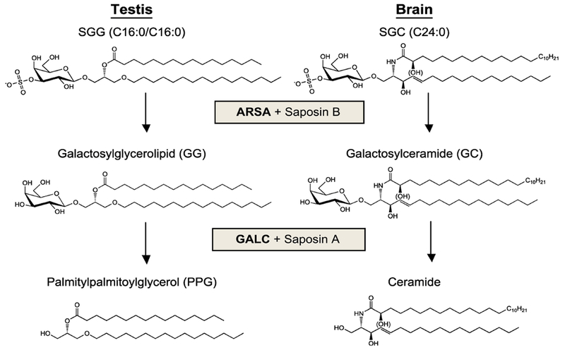
Sulfogalactosylglycerolipid (SGG, aka seminolipid) is selectively synthesized in high amounts in mammalian testicular germ cells (TGCs). SGG is an ordered lipid and directly involved in cell adhesion. SGG is indispensable for spermatogenesis, a process that greatly depends on interaction between Sertoli cells and TGCs. Spermatogenesis is disrupted in mice null for Cgt and Cst, encoding two enzymes essential for SGG biosynthesis. Sperm surface SGG also plays roles in fertilization. All of these results indicate the significance of SGG in male reproduction. SGG homeostasis is also important in male fertility. Approximately 50% of TGCs become apoptotic and phagocytosed by Sertoli cells. SGG in apoptotic remnants needs to be degraded by Sertoli lysosomal enzymes to the lipid backbone. Failure in this event leads to a lysosomal storage disorder and sub-functionality of Sertoli cells, including their support for TGC development, and consequently subfertility. Significantly, both biosynthesis and degradation pathways of the galactosylsulfate head group of SGG are the same as those of sulfogalactosylceramide (SGC), a structurally related sulfoglycolipid important for brain functions. If subfertility in males with gene mutations in SGG/SGC metabolism pathways manifests prior to neurological disorder, sperm SGG levels might be used as a reporting/predicting index of the neurological status.

Keywords: Lipid rafts; Lipidomics; Male fertility; Male reproduction; Mass spectrometry; Seminolipid; Sulfogalactosylglycerolipid.


Conflict of interest statement

Conflicts of interest

None

Figures

Figure 1.
Figure 1.. Structures of SGG and SGC.
Although the two sulfoglycolipids have different lipid backbones, alkylated glycerol for SGG and sphingosine for SGC, their overall conformation is similar to each other. Both SGG and SGC have the same galactose-3’-sulfate head group and each has two hydrocarbon chains, which insert into the lipid bilayers. For SGG, an alkyl and an acyl chain are present at the sn-1 and sn-2 position of glycerol, respectively. For SGC, an acyl chain is N-linked to the long chain base d18:1 sphingosine. The trans double bond in the sphingosine d18:1 still allows tight chain packing as in a saturated hydrocarbon chain. As shown here, C16:0/C16:0 SGG is a prevalent molecular species in male germ cells, whereas (hydroxylated) C24:0 SGC is one of the abundant molecular species of SGC found in the brain and other tissues.
Figure 2.
Figure 2.. SGG quantification by multiple reaction monitoring (MRM) in triple quadrupoles (Q).
Upper panel: Schematic representation of the multiple reaction monitoring experiment in which parent ions (P in the lower panel) of interest (in this case the ions at m/z 798 and 795) are sequentially selected in the first mass analyzer (Q1). These ions are allowed to collide with gas molecules in the collision cell (Q2); the emerging ions are separated in the second mass analyzer (Q3), and their relative intensity is recorded by the detector. The arrows indicate the sites at which fragmentation takes place, and the proposed structures of the fragment are shown. Lower panel: Fragment ion mass spectra of deuterated SGG internal standard (Top) and natural (unlabeled) SGG (Bottom). The deuterated SGG contains a terminal C2H3 (CD3) moiety instead of CH3 in the natural SGG form, thus making the m/z values of the parent, and any fragment ions containing the CD3 moiety, 3 Daltons heavier than the corresponding ions from the natural SGG form. Both observed and calculated ion masses are shown as the integer values. The mass spectra presented are taken from Franchini et al. [53].
Figure 3.
Figure 3.
A: The seminiferous epithelium (SE), the site of spermatogenesis. Somatic Sertoli cells span through the whole thickness of the SE. Between adjacent Sertoli cells towards the basement membrane, there exist tight junctions (TJs) and basal ectoplasmic specializations (ESs), which together form the blood-testis barrier. Both TJs and ESs are bordered by actin filaments with TJ and ES proteins interconnecting between the two adjacent Sertoli cells, as shown in the top inset 1 (drawn based on the previous publication [239]). TJ proteins include occludin, ZO1, ZO2 and ZO3, whereas nectin/afadin, cadherin/catenin and γ-catenin constitute ES junctions. Spermatogonia undergoing mitosis and preleptotene spermatocytes are localized underneath these junctions, whereas other developing testicular germ cells (TGCs) in meiotic and morphogenetic phases of development are in the adluminal compartment above. During the course of their development, TGCs move towards the lumen, with the most developed (i.e., spermatids) being closest to the lumen. Each successive layer of TGCs can be referred to as a distinct generation arising from amplifying progenitor spermatogonia at precisely timed intervals depending on the species (16 days in human, 12 days in mice). Note that secondary spermatocytes are short lived and therefore are not always present in the SE. Finally, elongating/elongated spermatid heads are inserted into the recesses of the Sertoli cell apical membrane. Apical ESs in Sertoli cells surround these elongated spermatids, holding them in place. As part of maturation to become testicular spermatozoa, the residual bodies (extra cytoplasm) are shed from the elongated spermatids and phagocytosed by Sertoli cells (see the top inset 2). Testicular spermatozoa are then released into the lumen. In mammals, there are two major forms of sperm heads, falciform and spatulate (see bottom inset). Note that this is a simplified drawing, which does not include all types of primary spermatocytes and various steps of spermatids. Information on the former aspect is described in B. More information on the second aspect as well as information on how various TGCs associate with each other in SE layers during different time intervals of spermatogenesis can be obtained from the recent review [11]. B: Steps in spermatogenesis. Differentiated spermatogonia undergo mitosis divisions and transform in a number of steps to become preleptotene primary spermatocytes, in which DNA replication takes place. With syntheses of more macromolecules, the more developed primary spermatocytes become enlarged and their various stages are classified by the increasing chromatin condensation (i.e., leptotene, zygotene and pachytene spermatocytes). Pachytene spermatocytes (distributed at the highest percentage among all primary spermatocytes in the seminiferous epithelium) then enter the first meiotic division to generate haploid secondary spermatocytes, which in turn undergo the second meiotic division to produce haploid round spermatids with DNA content being one half of that of secondary spermatocytes. Finally, round spermatids are differentiated through a number of steps to become elongated spermatids and then testicular spermatozoa. Therefore, one pachytene spermatocyte should generate four testicular spermatozoa. However, apoptosis usually occurs en route [96], thus yielding a lower number of spermatozoa than expected. It takes ~45 days in mice for one Type A spermatogonium to develop into a testicular spermatozoon. Drawing was adapted from Bellve el al. [72].
Figure 4.
Figure 4.. SGG levels in mouse testicular germ cells and sperm
Purified populations of primary spermatocytes, round spermatids and caudal epididymal/vas deferens sperm isolated from CD-I male mice (kindly provided by Dr. Stuart Moss, University of Pennsylvania) were subjected to lipid extraction and SGG quantification by ESI-MS/MS MRM. The data were expressed as mean ± S.D. of results from triplicate experiments and are taken from K Kongmanas’ Ph.D. thesis, 2015, University of Ottawa, which has been published on line (https://ruor.uottawa.ca/handle/10393/32509).
Figure 5.
Figure 5.. Presence of SGG in Sertoli cells and testicular germ cells in mice of different ages.
A: Mass spectra and reconstructed ion images (insets) from MS imaging showing the absence of SGG (m/z 795) in Sertoli cells (top panel) and spermatogonia cells (middle panel), both isolated from 5-day-old mice, but its presence in TGCs of 20-day-old mice (bottom panel). B: HPTLC showing the presence of SGG in Sertoli cells of 20-day-old mice but not in those of the 5-day-old animals. Lipids extracted from 2 million Sertoli cells isolated from 5-day-old or 20-day-old mice were loaded onto a HPTLC plate, and stained with orcinol solution which specifically reacted with glycolipids to yield purple bands. SGG was found as the major glycolipid in the Sertoli cells of 20-day-old mice (where the first round of spermatogenesis has initiated) but it was not detectable in Sertoli cells of neonatal 5-day-old mice. C: Panel a: Mass spectra from precursor ion scanning of m/z 97 (sulfate group) showing the presence of SGG (m/z 795) as the major sulfolipid in Sertoli cells isolated from 20-day-old (top) and 10-week-old (bottom) mice. Panel b: Table showing the amounts of SGG in Sertoli cells of 20-day-old and 10-week-old mice quantified by ESI-MS/MS-MRM analysis. Data are expressed as mean ± S.D. of triplicate results. All data presented are taken from K Kongmanas’ Ph.D. thesis, 2015, University of Ottawa, which has been published on line (https://mor.uottawa.ca/handle/10393/32509).
Figure 6.
Figure 6.. Biosynthesis pathway of the galactosylsulfate head group of SGG and SGC.
The two sulfogalactolipids utilize the same biosynthesis pathway for their head group, which was discerned mainly from studies on Cgt and Cst null mice (see text for more details). CGT: UDP-galactose:ceramide galactosyltransferase; CST: PAPS: cerebroside sulfotransferase. PAPS: 3’-phosphoadenosine-5’-phosphosulfate.
Figure 7.
Figure 7.. Preparation of Sertoli cell plasma membrane proteins with specific affinity to SGG.
Plasma membrane proteins were extracted from the primary culture of Sertoli cells by 2% octylglucoside in PBS. Following removal of the detergent by dialysis, the extracted proteins were incubated (37°C, 1 h) with multilamellar liposomes of DPPC/cholesterol (molar ratio, 2:1) in PBS. DPPC/cholesterol liposomes with bound proteins were then pelleted by ultracentrifugation. The supernatant obtained was further incubated (37°C, 1 h) with multilamellar liposomes of SGG/cholesterol (molar ratio, 2:1) in PBS, and SGG/cholesterol liposomes with bound proteins were likewise pelleted by ultracentrifugation. Proteins bound to DPPC/cholesterol liposomes and SGG/cholesterol liposomes were then solubilized in Laemmli’s sample buffer and subjected to SDS-PAGE. Both DPPC and SGG multilamellar liposomes were prepared as previously described Attar et al. [38].
Figure 8.
Figure 8.
A: Identification of Sertoli cell plasma membrane proteins with SGG affinity. Proteins bound to DPPC and SGG liposomes prepared as described in the legend of Figure 7 were subjected to SDS-PAGE followed by silver staining. Since the amounts of DPPC bound proteins were much higher than those of SGG bound proteins in one preparation, only 1/10 by weight of DPPC bound proteins were loaded as compared with SGG bound proteins. Comparison of the stained gel profile indicates that protein bands of 91, 86, 39, 30 and 20 kDa were present specifically in SGG bound protein samples. The 91 and 86 kDa bands were excised from both SGG bound and DPPC bound protein lanes and in-gel digested with trypsin. Tryptic peptides obtained were subjected to linear ion trap based Fourier transform MS analyses. Identities of proteins (UniProt nomenclature) present in the two excised gel bands were shown in the table along with their average ion scores in the SGG bound and DPPC bound protein samples. B: Immunoblotting of erzin (E), radixin (R) and moesin (M) proteins. The whole Sertoli cell plasma membrane extract (IN) was loaded as a positive control. SGG and DPPC denote proteins that were bound to SGG and DPPC liposomes, respectively. Note that ERM proteins were specifically present in the SGG bound protein sample. All data presented are unpublished and obtained from the study performed by B Doyle, K Kongmanas and N Tanphaichitr, University of Ottawa, and J Whitelegge, UCLA.
Figure 9.
Figure 9.
A: Steps in sperm maturation and sperm-egg interaction. Testicular sperm cannot fertilize eggs. They acquire fertilizing ability in a stepwise manner. 1. They undergo “maturation” during their transit through the proximal part of the epididymis and during their storage in the distal (cauda) epididymis. Epididymal sperm acquire forward movement. As well, a number of egg binding proteins present in the epididymal lumen deposit onto their head surface. 2. Sperm gain full fertilizing ability in the female reproductive tract through the so-called “capacitation” process, which involves cholesterol efflux, leading to an increase in membrane fluidity and signal transduction. As a result, their motility becomes hyperactivated with whiplash patterns. Sperm protein tyrosine phosphorylation is distinctively increased and ZP binding proteins are fully exposed on the sperm head surface. Despite cholesterol efflux, sperm lipid rafts increase in amount following capacitation. 3. During capacitated sperm movement through the cumulus cell layers surrounding mature eggs, acrosomal exocytosis is usually initiated in mice. Released acrosomal hydrolytic enzymes likely digest the egg vestments facilitating sperm to swim to the ZP. Sperm-ZP interaction then takes place in a species-specific manner, involving a number of ZP binding proteins present on the sperm surface as well as in the acrosomal matrix. 4. Finally, acrosome reacted sperm that have penetrated through the ZP layer bind to the egg plasma membrane. Following sperm-egg plasma membrane interaction, one acrosome reacted sperm enters the egg cytoplasm, signifying that fertilization has occurred. B: Presence of SGG in both acrosome intact and reacted sperm. SGG has direct affinity for the ZP [6]. Therefore, it is involved in sperm-ZP interaction according to the model presented in A, step 3. The role of sperm SGG in sperm-ZP binding also fits into the earlier model describing that only acrosome intact sperm bind to the ZP [167].
Figure 10.
Figure 10.. Degradation pathway of the galactosylsulfate head group of SGG and SGC.
The degradation pathway of the galactosylsulfate head group of SGC in the brain was discerned mainly from samples of humans with natural mutations of the catabolic enzymes, ARSA and GALC, and their co-enzymes, SAP-B and SAP-A, respectively. The degradation pathway of SGG was subsequently revealed through studies employing Arsa knockout mice and twitcher mice (lacking GALC due to gene mutation by ENU).
Figure 11.
Figure 11.. Increased levels of SGC in the brain of Arsa knockout mice relative to age-matched wild type mice.
Lipids were extracted from brain tissues of wild type and Arsa null mice of 5 months and 8 months of age and subjected to ESI-MS/MS-MRM of SGC of various molecular species (with m/z values shown in the top right comer). Data are expressed as means ± SDs from three replicate experiments. * denotes a statistical difference of the level of each SGC molecular species in the brain between Arsa null mice and age-matched wild types. Note the increases in SGC levels in the knockout mice. Although there was no statistical difference of the levels of C24:1 and C24:0 SGC between 8-month old Arsa knockout mice and the wild type counterparts, the trend showing higher levels of these SGC molecular species in the knockout mice was obvious. Data presented are taken from K Kongmanas’ Ph.D. thesis, 2015, University of Ottawa, which has been published on line (https://ruor.uottawa.ca/handle/10393/325091).
Figure 12.
Figure 12.. Metabolisms of SGG in seminiferous tubules: interrelationship between testicular germ cells and Sertoli cells.
Top panel: In normal testes, Sertoli cells degrade SGG in phagocytosed residual bodies and apoptotic TGC remnants to GG, and then to PPG by lysosomal ARSA and GALC, respectively Note that in Sertoli cells of wild type animals, phagocytosed apoptotic germ cells or remnants thereof are not evident indicating the rapid pace of their degradation by lysosomal enzymes. Likewise, late residual bodies disappear in due time. PPG produced from the SGG degradation pathway may be shuttled to developing pachytene spermatocytes (pSc) for new SGG synthesis, although pSc themselves would also have the ability to synthesize PPG de novo. See box for the SGG degradation and synthesis pathways. Bottom left panel: In 5-month old Arsa knockout mice, SGG is accumulated in Sertoli cells due to the lack of ARSA, although these higher levels of intracellular SGG have not yet reached the cytotoxic threshold. With no PPG produced in Sertoli cells for recycling, pSc would likely have to increase the de novo synthesis of PPG, and therefore the amounts of SGG in TGCs of these knockout mice are the same as those in age-matched wild type males. Bottom right panel: In 8-month old Arsa null mice, the much higher levels of accumulated SGG exert cytotoxicity to Sertoli cells, which then become smaller in size with their nuclei (N) displaced to be on top of the basement membrane. Sertoli cells in these aging Arsa null mice presumably are much less functional having reduced support for the development of TGCs. pSc in turn synthesize PPG at reduced rates and the subsequent levels of SGG in TGCs in these knockout mice are only 50% of the corresponding wild type levels. Also, the shaping of the elongated spermatid head, as regulated by Sertoli cells, is aberrant, resulting in production of sperm with abnormal morphology. BTB: blood-testis barrier, Ly: lysosome, Rb: residual body, SG: spermatogonium, lSc: leptotene spermatocyte, RS: round spermatid, ES: elongated spermatid. Adapted from Xu et al. [16].

References

    1. Ishizuka I, Suzuki M, Yamakawa T, Isolation and characterization of a novel sulfoglycolipid, seminolipid, from boar testis and spermatozoa, J. Biochem 73 (1973) 77–87. - PubMed
    1. Tanphaichitr N, Bou Khalil M, Weerachatyanukul W, Kates M, Xu H, Carmona E, Attar M, Carrier D, Physiological and biophysical properties of male germ cell sulfogalactosylglycerolipid, in: De Vriese S (Ed.), Lipid Metabolism and Male Fertility, AOCS Press, Champaign, IL, 2003, pp. 125–148.
    1. Tanphaichitr N, Carmona E, Bou Khalil M, Xu H, Berger T, Gerton GL, New insights into sperm-zona pellucida interaction: involvement of sperm lipid rafts, Front. Biosci 12 (2007) 1748–1766. - PubMed
    1. Tanphaichitr N, Faull KF, Yaghoubian A, Xu H, Lipid rafts and sulfogalactosylglycerolipid (SGG) in sperm functions: consensus and controversy, Trends Glycosci. Glycotech 19(106) (2007) 67–83.
    1. Rajendran L, Simons K, Lipid rafts and membrane dynamics, J. Cell Sci 118(Pt 6) (2005) 1099–1102. - PubMed

Publication types