. 2018 Dec:120:1-11.
doi: 10.1016/j.nbd.2018.08.013. Epub 2018 Aug 24.

The glycoprotein GPNMB is selectively elevated in the substantia nigra of Parkinson's disease patients and increases after lysosomal stress

Affiliations

The glycoprotein GPNMB is selectively elevated in the substantia nigra of Parkinson's disease patients and increases after lysosomal stress

Elizabeth B Moloney et al. Neurobiol Dis. 2018 Dec.

Abstract

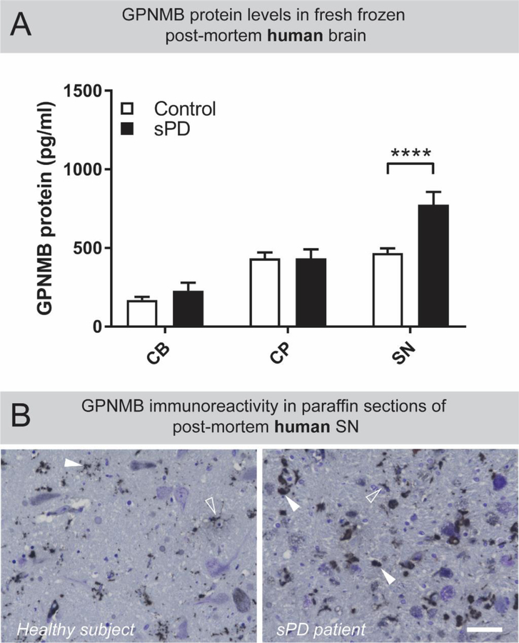
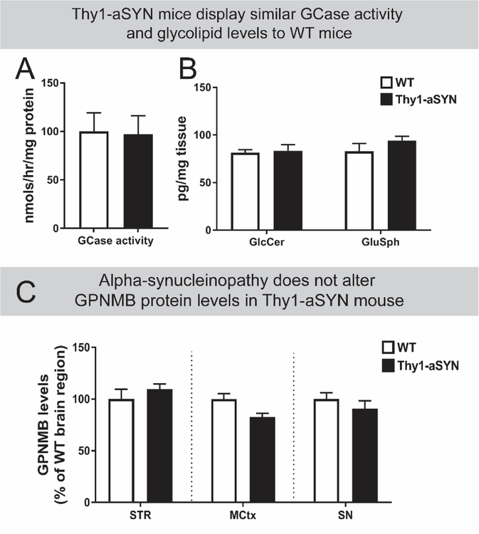
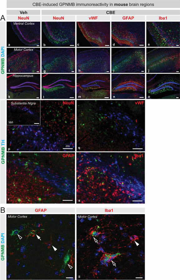
GPNMB is a glycoprotein observed upon tissue damage and inflammation and is associated with astrocytes, microglia, and macrophages. Gene variations in GPNMB are linked with Parkinson's disease (PD) risk, and changes in protein levels of GPNMB have been found in lysosomal storage disorders, including Gaucher's disease with glucocerebrosidase (GCase) deficiency. In the current study, GPNMB increases were seen in the substantia nigra (SN) of PD patients compared to age-matched controls. Such PD patients have a decrease in GCase activity and corresponding elevation of glycosphingolipids in the SN (Rocha et al., 2015a). Interestingly, transgenic mice modelling synucleinopathy did not show GPNMB elevations or altered GCase activity levels compared to wild-type mice. However, upon CBE-induced GCase lysosomal dysfunction with elevated glycosphingolipids in wild-type mice, there were similar changes in GPNMB levels in the brain as seen in PD patient brains. These results indicate that GPNMB levels do not depend on alpha-synuclein load per se but relate directly to the lipidopathy changes induced by CBE-mediated GCase inhibition. The experimental modelling of elevating glycolipids resulted in GPNMB elevations with glial activation in several brain regions in mice. This is the first demonstration of region-specific elevations of GPNMB protein in Parkinson's disease. The presence of GPNMB in PD patient substantia nigra, the induction of GPNMB after experimental glycosphingolipid increases, but not with pure alpha-synucleinopathy, point towards the potential for primary lipid-induced degeneration in PD.

Keywords: GPNMB; Glucocerebrosidase; Lipidopathy; Lysosome; Parkinson's disease; Post-mortem.


Figures

Figure 1:
Figure 1:. GPNMB is uniquely elevated in the substantia nigra of sporadic PD patients
(A) Tissue homogenates from post-mortem cerebellum (CB) caudate and putamen (CP) and substantia nigra (SN) from healthy subject controls (n=31) and sporadic PD patients (n=25) were prepared. Levels of GPNMB in these brain regions were determined by ELISA. Results are means ±SEM, **** p <0.0001 (two-way ANOVA, with Sidak’s multiple comparison post-hoc test). (B) DAB-based immunostaining revealed that GPNMB-positive puncta exist throughout the human SN (white arrowheads) but are larger and more numerous in sporadic PD patients compared to healthy subjects. Additionally, some GPNMB-positive immunoreactivity produced a cell-like staining pattern (open arrowheads) in both healthy subjects and sporadic PD patients. Scale bar is 50µm.
Figure 2:
Figure 2:. GPNMB is elevated in mouse brain following complete pharmacological inhibition of glucocerebrosidase activity.
Tissue homogenates of freshly dissected striatum (STR), cerebellum (CB), motor cortex (MCtx), hippocampus (HP) and substantia nigra (SN) from mice receiving daily (for 28 days) i.p. injections of DMSO (0.1%, n=4–6) or CBE (100mg/kg, n=3–6) were prepared. (A) Glucocerebrosidase activity levels were determined using the artificial substrate 4-methylumbelliferyl-β-D-glucopyranoside (4-MU-Glc). GCase activity levels were significantly diminished in all brain regions in CBE-treated mice. (B) Levels of GPNMB across these brain regions were determined by ELISA, with significantly elevated GPNMB measured in the MCtx, HP, and SN of CBE-treated mice. GPNMB levels in the STR and CB were not affected by CBE-treatment. For panels A and B, results are means ±SEM, ** p <0.01, **** p <0.0001 (two-way ANOVA, with Sidak’s multiple comparison post-hoc test).
Figure 3:
Figure 3:. GPNMB levels in brain regions of mice modelling alpha-synucleinopathy are not different compared to age-matched WT mice.
Whole brain homogenates from age-matched wild-type (WT; n=6) or Thy1 alpha-synuclein overexpressing (Thy1-aSYN; n=6) mice display similar levels of (A) glucocerebrosidase (GCase) activity and (B) glycosphingolipids such as glucosylceramide (GlcCer) and glucosphingosine (GluSph). Results are mean ±SEM. (C) GPNMB levels in striatum (STR), motor cortex (MCtx), and substantia nigra (SN) motor cortex homogenates from age-matched WT (n=3–6) and Thy1-aSYN mice (n=6–9) were measured by ELISA and show that transgenic animals display similar GPNMB levels to WT mice in these brain regions. Results are mean normalized to corresponding WT brain region ±SEM. No significant differences were observed with statistical tests (Panel A: unpaired two-tailed Students T-test; Panels B and C: two-way ANOVA).
Figure 4:
Figure 4:. GPNMB is associated with glial cells in mouse brain following pharmacological inhibition of glucocerebrosidase activity.
(A) Fluorescent immunostaining in ventral cortex (a–e), motor cortex (f–j), and hippocampus (k–o) for GPNMB (green), DAPI (blue), and various cell-specific markers in red (NeuN, vWF, Iba1 and GFAP) illustrate that GPNMB immunoreactivity is elevated following CBE-treatment (b–e, g–j, l–o) compared to vehicle-treatment (a, f, k). A triple immunostaining was performed to visualize TH-positive cells (blue), GPNMB (green) and cell-specific markers (red): NeuN (p), vWF (q) GFAP (r), and Iba1 (s) in the substantia nigra (p–s), which also depicts that GPNMB immunoreactivity is increased upon CBE-treatment (p – s) compared to vehicle treatment (inset in p). (B) High magnification confocal microscopy images illustrate the variety of staining patterns obtained for GPNMB (green) in and around astrocytes (i; GFAP, red) or microglia (ii; Iba1, red). GPNMB immunoreactivity is found to be associated with GFAP-positive processes (closed arrow in top panel) or surrounded by Iba1-positive processes (open arrows in bottom panel). GPNMB-positive cell-like structures that are not GFAP-positive are also observed (open arrowheads). GFAP-positive astrocytes, or Iba1-positive microglia that are negative for GPNMB are also present (closed arrowheads). Scale bars in panel A = 100µm. Scale bars in panel B = 10µm.

References

    1. Aflaki E, Westbroek W, Sidransky E. The Complicated Relationship between Gaucher Disease and Parkinsonism: Insights from a Rare Disease. Neuron 2017; 93: 737–746.Available from: http://www.ncbi.nlm.nih.gov/pubmed/28231462 - PMC - PubMed
    1. Ahn JH, Lee Y, Jeon C, Lee S-JJ, Lee B-HH, Choi KD, et al. Identification of the genes differentially expressed in human dendritic cell subsets by cDNA subtraction and microarray analysis. Blood 2002; 100: 1742–54.Available from: http://www.ncbi.nlm.nih.gov/pubmed/12176896 - PubMed
    1. Batta G, Soltész L, Kovács T, Bozó T, Mészár Z, Kellermayer M, et al. Alterations in the properties of the cell membrane due to glycosphingolipid accumulation in a model of Gaucher disease. Sci. Rep 2018; 8: 157.Available from: http://www.ncbi.nlm.nih.gov/pubmed/29317695 - PMC - PubMed
    1. Blanz J, Saftig P. Parkinson’s disease: acid-glucocerebrosidase activity and alpha-synuclein clearance. J. Neurochem 2016; 139: 198–215.Available from: http://www.ncbi.nlm.nih.gov/pubmed/26860955 - PubMed
    1. Bossi SR, Simpson JR, Isacson O. Age dependence of striatal neuronal death caused by mitochondrial dysfunction. Neuroreport 1993; 4: 73–76.Available from: http://www.ncbi.nlm.nih.gov/pubmed/8453041 - PubMed

Publication types

Substances