. 2018 Aug 24;7(9):116.
doi: 10.3390/cells7090116.

Cytotoxic Constituents from the Sclerotia of Poria cocos against Human Lung Adenocarcinoma Cells by Inducing Mitochondrial Apoptosis

Affiliations

Cytotoxic Constituents from the Sclerotia of Poria cocos against Human Lung Adenocarcinoma Cells by Inducing Mitochondrial Apoptosis

Seulah Lee et al. Cells. .

Abstract

Previous studies have revealed the antitumor potential of Poria cocos Wolf against a broad spectrum of cancers. However, the biological activity of P. cocos against lung cancer, which is known as the leading cause of cancer mortality worldwide, and its underlying chemical and molecular basis, remain to be investigated. We aimed to evaluate the in vitro cytotoxicity of P. cocos toward human lung adenocarcinoma cells with different p53 statuses, to identify the bioactive constituents of P. cocos, and explicate the molecular mechanisms underlying the cytotoxicity of these constituents in human lung adenocarcinoma cells. An EtOH extract of the sclerotia of P. cocos exhibited cytotoxicity toward four human lung cancer cell lines: A549, H1264, H1299, and Calu-6, regardless of their p53 status. Chemical investigation of the extract resulted in the isolation of two triterpenoids, dehydroeburicoic acid monoacetate (1) and acetyl eburicoic acid (4); a sterol, 9,11-dehydroergosterol peroxide (2); and a diterpenoid, dehydroabietic acid (3). All of the isolated compounds were cytotoxic to the lung adenocarcinoma cell lines, exhibiting IC50 values ranging from 63.6 μM to 171.0 μM at 48 h of treatment. The cytotoxicity of the extract and the isolated compounds were found to be mediated by apoptosis, and accompanied by elevated Bax expression and/or Bcl-2 phosphorylation along with caspase-3 activation. Our data demonstrate that the sclerotium of P. cocos and its four bioactive constituents (14) exert cytotoxicity against human lung adenocarcinoma cells, regardless of their p53 status, by inducing apoptosis associated with mitochondrial perturbation, and proposing the potential to employ P. cocos in the treatment of lung cancer.

Keywords: Poria cocos; apoptosis; cytotoxicity; diterpenoid; lung cancer; polyporaceae; sterol; triterpenoid.


Conflict of interest statement

The authors declare no conflict of interest.

Figures

Figure 1
Figure 1
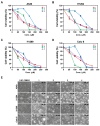
The EtOH extract of the sclerotia of P. cocos reduces cell viability in human lung adenocarcinoma cells, regardless of the p53 status. (A) Cell viability assessed with the WST-1 assay in four human lung adenocarcinoma cell lines, A549, H1264, H1299, and Calu-6, 48 h after treatment with the EtOH extract at the indicated concentrations. (B) Representative bright-field images (200× total magnification) of human lung adenocarcinoma cells treated with the EtOH extract at the indicated concentrations for 48 h. Magnified regions are shown as insets. Data are presented as means ± SEMs. Scale bar: 100 μm.
Figure 2
Figure 2
The EtOH extract of the sclerotia of P. cocos induces apoptosis in human lung adenocarcinoma cells. (AD) Representative fluorescence images (400× total magnification) of TUNEL (green) and 4′,6-diamidino-2-phenylindole (DAPI) (blue) staining (left), and quantitation of TUNEL-positive cells (right), in A549 (A), H1264 (B), H1299 (C), and Calu-6 (D) cells treated with 400 μg/mL of the EtOH extract or 0.4% DMSO as a vehicle control for 48 h. Data are presented as means ± SEMs. Scale bar: 50 μm. ** p < 0.01.
Figure 3
Figure 3
Structures of compounds (14) isolated from the sclerotia of P. cocos.
Figure 4
Figure 4
Cytotoxicity of compounds isolated from the EtOH extract of the sclerotia of P. cocos toward human lung adenocarcinoma cells. (AD) Cell viability in A549 (A), H1264 (B), H1299 (C), and Calu-6 (D) cells, assessed with the WST-1 assay 48 h after treatment with the isolated compounds (14) at the indicated concentrations. (E) Representative bright-field images (200× total magnification) of human lung adenocarcinoma cells treated with the isolates at the indicated concentrations or 0.8% DMSO as a vehicle control. Data are presented as means ± SEMs. Scale bar: 100 μm.
Figure 5
Figure 5
Pro-apoptotic effects of isolated compounds 14 in human lung adenocarcinoma cells. (AD) Representative fluorescence images (400× total magnification) of TUNEL (green) and DAPI (blue) staining (left), and quantitation of TUNEL-positive cells (right), in A549 (A), H1264 (B), H1299 (C), and Calu-6 (D) cells treated with the compounds (14) or DMSO (DM) as a vehicle control at the indicated concentrations for 48 h. Data are presented as means ± SEMs. Scale bar: 50 μm. ** p < 0.01.
Figure 6
Figure 6
The EtOH extract of the sclerotia of P. cocos and isolated compounds 14 activate caspase-3 and alter Bax and Bcl-2 proteins in human lung adenocarcinoma cells. (A,B) Calu-6 cells were treated for 48 h with 400 μg/mL of the EtOH extract (A) and with 100 μM, 75 μM, 150 μM, and 150 μM of compounds 1, 2, 3, and 4, respectively (B). Cells treated with 1 μM of doxorubicin (Doxo) and 0.4 or 0.75% DMSO served as positive and vehicle controls, respectively. Whole-cell lysates were then prepared and probed for caspase-3, cleaved caspase-3, PARP, Bax, Bcl-2, and β-actin (a loading control).

References

    1. Fitzmaurice C., Allen C., Barber R.M., Barregard L., Bhutta Z.A., Brenner H., Dicker D.J., Chimed-Orchir O., Dandona R., Dandona L., et al. Global, regional, and national cancer incidence, mortality, years of life lost, years lived with disability, and disability-adjusted life-years for 32 cancer groups, 1990 to 2015: A systematic analysis for the global burden of disease study. JAMA Oncol. 2017;3:524–548. - PMC - PubMed
    1. Jemal A., Siegel R., Xu J., Ward E. Cancer statistics. CA Cancer J. Clin. 2010;60:277–300. doi: 10.3322/caac.20073. - DOI - PubMed
    1. Wasser S.P. Current findings, future trends, and unsolved problems in studies of medicinal mushrooms. Appl. Microbiol. Biotechnol. 2011;89:1323–1332. doi: 10.1007/s00253-010-3067-4. - DOI - PubMed
    1. Paterson R.R., Lima N. Biomedical effects of mushrooms with emphasis on pure compounds. Biomed. J. 2014;37:357–368. doi: 10.4103/2319-4170.143502. - DOI - PubMed
    1. Maehara Y., Tsujitani S., Saeki H., Oki E., Yoshinaga K., Emi Y., Morita M., Kohnoe S., Kakeji Y., Yano T., et al. Biological mechanism and clinical effect of protein-bound polysaccharide K (KRESTIN®): Review of development and future perspectives. Surg. Today. 2012;42:8–28. doi: 10.1007/s00595-011-0075-7. - DOI - PMC - PubMed

LinkOut - more resources