Review
. 2018 Aug 25;18(9):2799.
doi: 10.3390/s18092799.

Breast Cancer Detection Using Infrared Thermal Imaging and a Deep Learning Model

Affiliations
Review

Breast Cancer Detection Using Infrared Thermal Imaging and a Deep Learning Model

Sebastien Jean Mambou et al. Sensors (Basel). .

Abstract

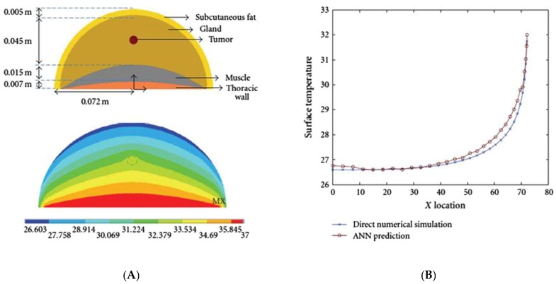
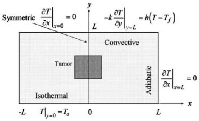
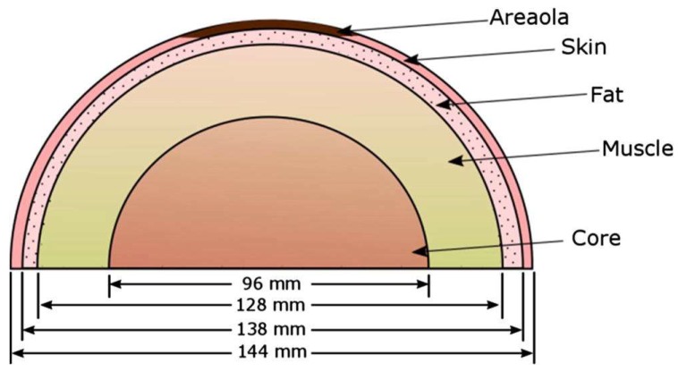
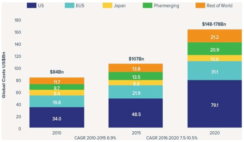
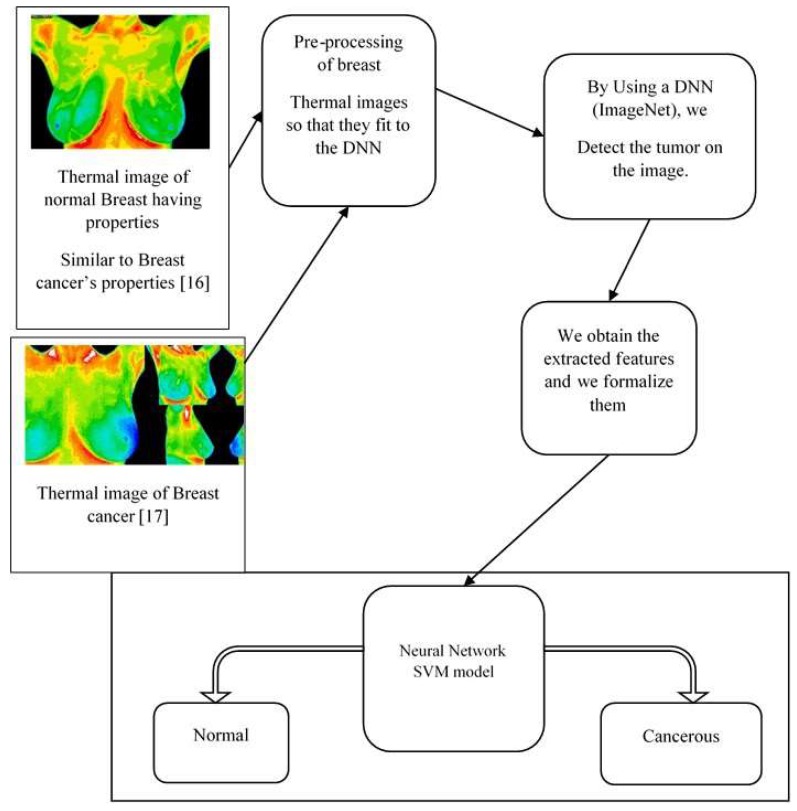
Women's breasts are susceptible to developing cancer; this is supported by a recent study from 2016 showing that 2.8 million women worldwide had already been diagnosed with breast cancer that year. The medical care of a patient with breast cancer is costly and, given the cost and value of the preservation of the health of the citizen, the prevention of breast cancer has become a priority in public health. Over the past 20 years several techniques have been proposed for this purpose, such as mammography, which is frequently used for breast cancer diagnosis. However, false positives of mammography can occur in which the patient is diagnosed positive by another technique. Additionally, the potential side effects of using mammography may encourage patients and physicians to look for other diagnostic techniques. Our review of the literature first explored infrared digital imaging, which assumes that a basic thermal comparison between a healthy breast and a breast with cancer always shows an increase in thermal activity in the precancerous tissues and the areas surrounding developing breast cancer. Furthermore, through our research, we realized that a Computer-Aided Diagnostic (CAD) undertaken through infrared image processing could not be achieved without a model such as the well-known hemispheric model. The novel contribution of this paper is the production of a comparative study of several breast cancer detection techniques using powerful computer vision techniques and deep learning models.

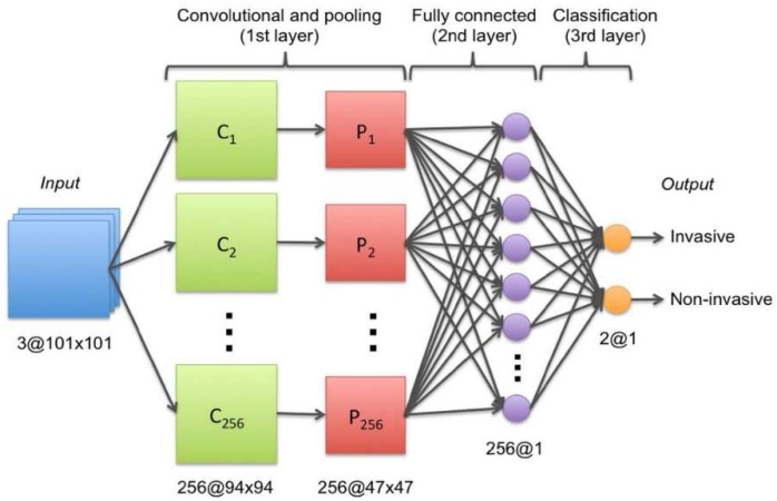
Keywords: DNN; RNN; SVM; breast; cancer; deep learning; detection; neural network; visual techniques.


Conflict of interest statement

The authors declare no conflict of interest.

Figures

Figure 1
Figure 1
ConvNet architecture used to determine the state of the cancer (invasive or non-invasive) [5].
Figure 2
Figure 2
Illustration of thermal diffusion in a conventional breast representation [22]. (A) The computational domain as a hemisphere semi-spherical. (B) Numerical simulation and surface temperatures.
Figure 3
Figure 3
The rectangular area that provided the first glimpse of predictive models that relate surface temperature to tumor size and location [23].
Figure 4
Figure 4
Hemispherical domain with non-concentric layers, which is a prevalent model because of its ability to reproduce the surface temperature [22,24].
Figure 5
Figure 5
The overview of the ICA process combined with automated post-processing, used to detect a tumor in the breast more effectively [28].
Figure 6
Figure 6
The process of extraction of the tumor area [28].
Figure 7
Figure 7
ICG fluorescence with a specific light wavelength (820 nm) in the near infrared spectrum [20].
Figure 8
Figure 8
A phase of breast warming [17].
Figure 9
Figure 9
Global costs and growth of oncology, 2010–2020 [33].
Figure 10
Figure 10
Illustration of our model [34].
Figure 11
Figure 11
Illustration of the increase in the accuracy over the number of iterations. The training and the validation become stable after 3900 training steps.
Figure 12
Figure 12
The entropy decreases during the training of our model, which can be considered a positive sign (the ambiguity reduces in our model).
Figure 13
Figure 13
The spatial distribution of our (retrained InceptionV3) model extracted feature representation shows how well features are grouped. In Red, we see elements classified as Healthy (For Healthy Breast), and In Blue, we see features organized as Sick (For Sick Breast or Breast with Cancer).
Figure 14
Figure 14
Confusion Matrix of our LinearSVC (SVM) shows how confident our classifier is for each prediction.
Figure 15
Figure 15
Precision-Recall of our LinearSVC result is a useful measure of the success of our prediction. In information retrieval, precision is a measure of result relevancy, while recall is a measure of how many truly relevant results are returned.
Figure 16
Figure 16
A receiver operating characteristic (ROC) curves of our prediction, where the top left corner of the plot can be interpreted as “ideal” point (a false positive rate of zero, and a true positive rate of one).
Figure 17
Figure 17
Our SVM architecture. The input Vector our context is filled by the features extracted at the feature layer of our Inception V3.
Figure 18
Figure 18
Given image (A) as input, our model classifies the image as “sick” with a confidence of 0.78.
Figure 19
Figure 19
Given image (B) as input, our model classifies the image as “healthy” with a confidence of 0.94.

References

    1. National Breast Cancer Foundation . Breast Cancer Facts. National Breast Cancer Foundation; Sydney, Australia: 2016.
    1. Dongola N. Mammography in Breast Cancer. [(accessed on 24 August 2018)]; Available online: https://emedicine.medscape.com/article/346529-overview.
    1. Hamidinekoo A., Denton E., Rampun A., Honnor K., Zwiggelaar R. Deep learning in mammography and breast histology, an overview and future trends. Med. Image Anal. 2018;47:45–67. doi: 10.1016/j.media.2018.03.006. - DOI - PubMed
    1. Kosus N., Kosus A., Duran M., Simavlı S., Turhan N. Comparison of standard mammography with Digital mammography and Digital infrared thermal imaging for breast cancer screening. J. Turk. Ger. Gynecol. Assoc. 2010;11:152–157. doi: 10.5152/jtgga.2010.24. - DOI - PMC - PubMed
    1. Li H., Weng J., Shi Y., Gu W., Mao Y., Wang Y., Liu W., Zhang J. An improved deep learning approach for detection of thyroid papillary cancer in ultrasound images. Sci. Rep. 2018;8 doi: 10.1038/s41598-01825005-7. - DOI - PMC - PubMed