. 2018 Aug 25;10(9):289.
doi: 10.3390/cancers10090289.

miRNA-205 Nanoformulation Sensitizes Prostate Cancer Cells to Chemotherapy

Affiliations

miRNA-205 Nanoformulation Sensitizes Prostate Cancer Cells to Chemotherapy

Prashanth K B Nagesh et al. Cancers (Basel). .

Abstract

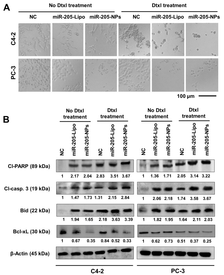
The therapeutic application of microRNA(s) in the field of cancer has generated significant attention in research. Previous studies have shown that miR-205 negatively regulates prostate cancer cell proliferation, metastasis, and drug resistance. However, the delivery of miR-205 is an unmet clinical need. Thus, the development of a viable nanoparticle platform to deliver miR-205 is highly sought. A novel magnetic nanoparticle (MNP)-based nanoplatform composed of an iron oxide core with poly(ethyleneimine)-poly(ethylene glycol) layer(s) was developed. An optimized nanoplatform composition was confirmed by examining the binding profiles of MNPs with miR-205 using agarose gel and fluorescence methods. The novel formulation was applied to prostate cancer cells for evaluating cellular uptake, miR-205 delivery, and anticancer, antimetastasis, and chemosensitization potentials against docetaxel treatment. The improved uptake and efficacy of formulations were studied with confocal imaging, flow cytometry, proliferation, clonogenicity, Western blot, q-RT-PCR, and chemosensitization assays. Our findings demonstrated that the miR-205 nanoplatform induces significant apoptosis and enhancing chemotherapeutic effects in prostate cancer cells. Overall, these study results provide a strong proof-of-concept for a novel nonviral-based nanoparticle protocol for effective microRNA delivery to prostate cancer cells.

Keywords: chemosensitivity and EMT; docetaxel; miR-205; prostate cancer.


Conflict of interest statement

The authors declare no conflict of interest.

Figures

Figure 1
Figure 1
Generation of miR-205-NPs formulations (A) Preparative approach and hypothetical structure of miR-205 nanoformulations. PEI-PEG: gray line belongs to PEI and dotted greens belong to PEG. miR-205 binds to PEI. (BC) Nanocomplexation assessment with miR-205. Fluorescence-based quenching study of fluorescein amidite (FAM)-labeled miRNA mimic MNP and MNP-MPEI-PEG (MPEI-PEG) nanoparticles. Note: MNP alone is also able to bind smaller amounts of miR-205, due to physical deposition on the nanoparticles but not due to binding. (D) Determination of nanocomplexation of miR-205 with MNP and MPEI-PEG through agarose gel electrophoresis. Data represent that 5 µg of nanocarrier is sufficient to hold 1 µg of miR-205 for delivery applications. (E) Evaluation of miR-205 binding with MNP and MPEI-PEG nanoparticles by circular dichroism (CD) spectral analysis.
Figure 2
Figure 2
miR-205 delivery characteristics of miR-205-NPs. (A,B) Hemolytic activity of MNP-PEI-PEG nanocarrier. Brightfield microscopy at 20× magnification was employed for capturing hemolytic characteristic of human red blood cells. Scale bar: 50 µm. Note: NPs are not toxic but lipofectamine is at the tested concentrations. Data indicate that nanocarrier is hemocompatible, unlike lipofectamine. (C) Dissociation studies of miR-205 in presence of poly(anion) (heparin). (D) miRNA-205 stability in the presence of 0–50% Fetal Bovine Serum (FBS) concentration. Equal amount of each sample was incubated with 10 μL of FBS at 37 °C for 24 h prior to gel electrophoresis. Note: “No NPs” lane did not show any band due to the absence of miRNA. (E) FAM-miRNA release profile from the FAM-miR through fluorescence spectral analysis at variable pH solutions (7.4, 6.5 and 3.5). The significance level was * p < 0.05. Each experiment has been repeated three times.
Figure 3
Figure 3
Cellular fate of miR-205-NPs formulation. (A) Cellular uptake of coumarin 6-labeled MPEI-PEG nanoformulation. Representative cellular internalized nanoparticles images were captured at 40× magnification using confocal microscopy. Green color arise from coumarin 6 in MPEI-PEG. Scale bar: 50 µm. (B) Quantitative measurement of FAM-miRNA mimic in cells treated with FAM-labeled miR-MPEI-PEG formulation. (C) Restoration of miR-205 through miR-205-NPs in PrCa cells. q-PCR gene expression studies reveals that treatments with miR-205-NPs reconstitutes the gene expression of miR-205. (D) miR-205-NPs influence cell growth in PrCa cells. Proliferation assays after miR-205 and miR-205-NPs transfection treatments in C4-2 and PC-3 cell lines using 3-(4,5-dimethylthiazol-2-yl)-5-(3-carboxymethoxyphenyl)-2-(4-sulfophenyl)-2H-tetrazolium, inner salt (MTS) assays. The significance level was * p < 0.05 with respect to NC/NPs. Each individual experiment has been repeated three times.
Figure 4
Figure 4
miR-205-NPs treatment chemosensitizes PrCa cells towards docetaxel therapy. (A) NC (No Dtxl), NPs, NC (non-targeting control), miR-205 naïve, miR-205-Lipo, and miR-205-NPs-treated PrCa cells (5 × 10³/well in 96-well plate) were treated with 0–25 nM Dtxl or respective control for 48 h and proliferation of cells was assessed by MTS assay. (B) NC, NPs, miR-205-Lipo, and miR-205-NPs-treated PrCa cells with 2.5 nM Dtxl. On day 14, cells were PBS-rinsed and stained with hematoxylin. Photographs of clonogenic pattern (in Multimage™ light cabinet) represent inhibition of clonogenic formation with miR-205-NPs formulation. Note: Figure 4B quantification was provided in Figure S3.
Figure 5
Figure 5
miR-205-NPs induce enhanced apoptotic potential of docetaxel in PrCa cells. (A) Representative microscopic images of various treatment groups after 24 h. Images were captured at 20× magnification. Scale bar: 100 µm. (B) Western blot analysis confirms the significant induction of apoptotic signaling proteins after the delivery of miR-205-NPs and Dtxl treatment. Cl-PARP and Cl-casp. 3 indicates cleaved PARP and cleaved caspase 3, respectively.
Figure 6
Figure 6
miR-205-NPs inhibits EMT signaling and sensitizes Dtxl treatment in PrCa cells. (A) miR-205-NPs treatment suppresses the migratory ability of C4-2 and PC-3 cells in presence/absence of the drug through Boyden chamber study. Images were captured at 20× magnification. Transwell assay with matrigel was performed to detect invasion activity of PrCa cells transfected with miR-205. Docetaxel treatments at 5 nM concentration. Scale bar: 200 µm. (B) Protein profiling studies of Control, miR-205 transfected and miR-205-NPs treated PrCa cells for 24 h for EMT signaling. Note: miR-205-L or miR-205-Lipo represents same and indicates to miR-205 transfected with lipofectamine.
Figure 7
Figure 7
miR-205-NPs effectively inhibit Pgp activity and facilitates Dtxl chemosensitization. (A,B) Rh123 Dye exclusion studies were performed in cells through morphological and flow cytometric methods. miR-205-NPs formulation facilitates Rh123, demonstrating its chemosensitizing potential. (C) miR-205-NPs inhibited MDR1/ABCB1 expression in both C4-2 and PC-3 cells and densitometry studies were calculated. (D) miR-205-NPs promote microtubule stabilization in PrCa cells. Except for non-targeting control (NC), 5 nM Dtxl was used in all treatment groups. Treatment period was 8 h. Images were captured at 40× magnification using confocal microscopy. Scale bar: 50 µm. Corrected total cell fluorescence (CTCF) values were calculated using ImageJ and showed as insets. The level of significance was * p < 0.05. Each individual experiment has been repeated three times.
Figure 8
Figure 8
Schematic representation of possible chemosensitization mechanism of action for miR-205-NPs formulation on prostate cancer cells.

References

    1. Siegel R., Naishadham D., Jemal A. Cancer statistics, 2013. CA Cancer J. Clin. 2013;63:11–30. doi: 10.3322/caac.21166. - DOI - PubMed
    1. Corcoran C., Rani S., O’Brien K., O’Neill A., Prencipe M., Sheikh R., Webb G., McDermott R., Watson W., Crown J., et al. Docetaxel-resistance in prostate cancer: Evaluating associated phenotypic changes and potential for resistance transfer via exosomes. PloS ONE. 2012;7:e50999. doi: 10.1371/journal.pone.0050999. - DOI - PMC - PubMed
    1. Hwang C. Overcoming docetaxel resistance in prostate cancer: A perspective review. Ther. Adv. Med. Oncol. 2012;4:329–340. doi: 10.1177/1758834012449685. - DOI - PMC - PubMed
    1. Cengiz E., Karaca B., Kucukzeybek Y., Gorumlu G., Gul M.K., Erten C., Atmaca H., Uzunoglu S., Karabulut B., Sanli U.A., et al. Overcoming drug resistance in hormone- and drug-refractory prostate cancer cell line, pc-3 by docetaxel and gossypol combination. Mol. Biol. Rep. 2010;37:1269–1277. doi: 10.1007/s11033-009-9501-y. - DOI - PubMed
    1. Pekarik V., Gumulec J., Masarik M., Kizek R., Adam V. Prostate cancer, miRNAs, metallothioneins and resistance to cytostatic drugs. Curr. Med. Chem. 2013;20:534–544. - PubMed

LinkOut - more resources