. 2018 Oct 1;201(7):2154-2164.
doi: 10.4049/jimmunol.1800124. Epub 2018 Aug 27.

Epidermal Growth Factor (EGF) Autocrine Activation of Human Platelets Promotes EGF Receptor-Dependent Oral Squamous Cell Carcinoma Invasion, Migration, and Epithelial Mesenchymal Transition

Affiliations

Epidermal Growth Factor (EGF) Autocrine Activation of Human Platelets Promotes EGF Receptor-Dependent Oral Squamous Cell Carcinoma Invasion, Migration, and Epithelial Mesenchymal Transition

Rui Chen et al. J Immunol. .

Abstract

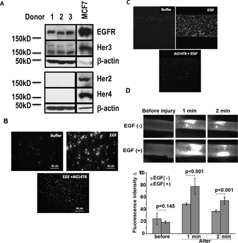
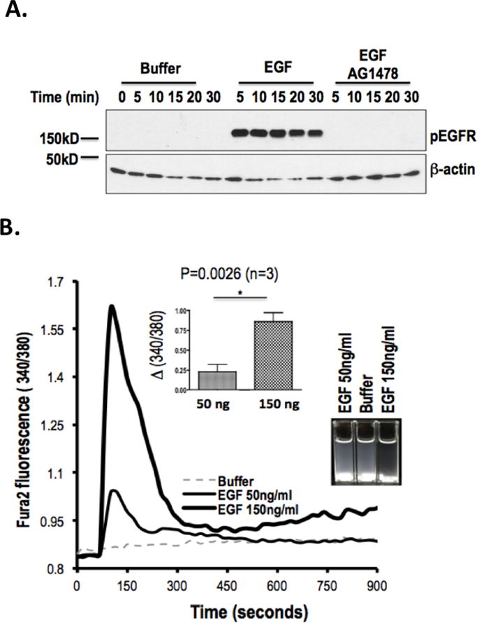
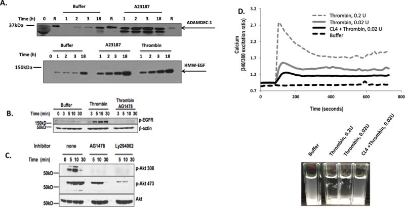
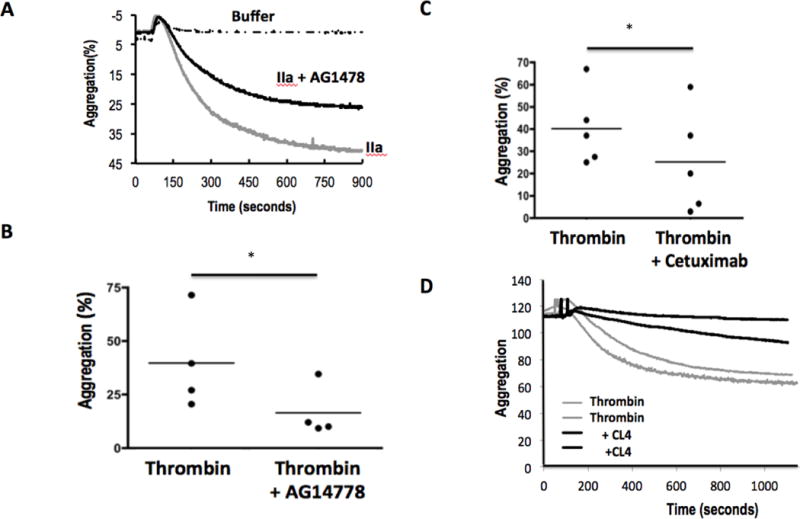
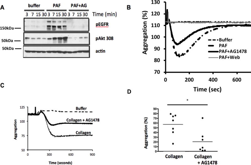
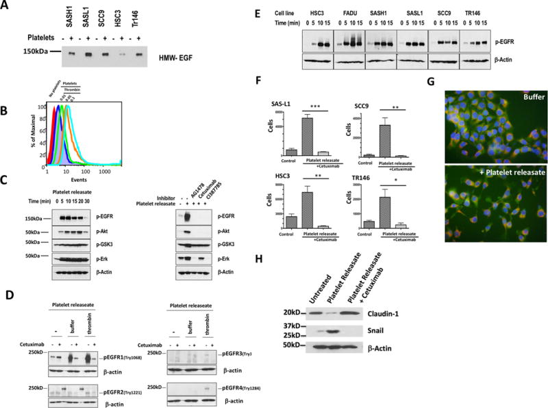
Activated platelets release functional, high m.w. epidermal growth factor (HMW-EGF). In this study, we show platelets also express epidermal growth factor (EGF) receptor (EGFR) protein, but not ErbB2 or ErbB4 coreceptors, and so might respond to HMW-EGF. We found HMW-EGF stimulated platelet EGFR autophosphorylation, PI3 kinase-dependent AKT phosphorylation, and a Ca2+ transient that were blocked by EGFR tyrosine kinase inhibition. Strong (thrombin) and weak (ADP, platelet-activating factor) G protein-coupled receptor agonists and non-G protein-coupled receptor collagen recruited EGFR tyrosine kinase activity that contributed to platelet activation because EGFR kinase inhibition reduced signal transduction and aggregation induced by each agonist. EGF stimulated ex vivo adhesion of platelets to collagen-coated microfluidic channels, whereas systemic EGF injection increased initial platelet deposition in FeCl3-damaged murine carotid arteries. EGFR signaling contributes to oral squamous cell carcinoma (OSCC) tumorigenesis, but the source of its ligand is not established. We find individual platelets were intercalated within OSCC tumors. A portion of these platelets expressed stimulation-dependent Bcl-3 and IL-1β and so had been activated. Stimulated platelets bound OSCC cells, and material released from stimulated platelets induced OSCC epithelial-mesenchymal transition and stimulated their migration and invasion through Matrigel barriers. Anti-EGF Ab or EGFR inhibitors abolished platelet-induced tumor cell phenotype transition, migration, and invasion; so the only factor released from activated platelets necessary for OSCC metastatic activity was HMW-EGF. These results establish HMW-EGF in platelet function and elucidate a previously unsuspected connection between activated platelets and tumorigenesis through rapid, and prolonged, autocrine-stimulated release of HMW-EGF by tumor-associated platelets.


Conflict of interest statement

Disclosures

The authors have no financial conflicts of interest.

Figures

Figure 1
Figure 1. Platelets express EGFR and respond to EGF
A. Platelets express EGFR. Proteins in the lysates of equivalent numbers of quiescent platelets were resolved by SDS-PAGE and immunoblotted for the EGFR family (Her2, Her3, Her4, EGFR) or β-actin as described in “Materials and Methods.” MCF7 cells are a positive control that express all EGFR family members. (n=3). B. EGF induces platelet adhesion. Platelets were labeled with the Ca++ sensitive dye Calcein-AM, treated or not with AG1478 to inhibit EGFR tyrosine kinase activity, and then incubated with EGF (200 ng/ml). These cells were then layered over albumin-coated glass slides, which suppress adhesion of quiescent platelets. Non-adherent cells were removed after 10 min by washing before Calcein fluorescence was imaged. (n=3) C. EGF stimulates platelet adhesion to collagen at high shear flow. Calcein-AM-labeled platelets were pulled through collagen-coated Cellix microfluidic channels at 63 Dynes with or without EGF and with or without AG14778 added at the initiation of flow. The assay was terminated after 5 min by pulling buffer through the channels and then imaging fluorescence at the chamber’s exit. (n=3) D. EGF augments intravascular thrombosis. Mice were injected with rhodamine 6G to fluorescently label platelets, with or without accompanying EGF (10 mg/kg), 10 min prior to ectopic application of FeCl3 to surgically exposed carotid arteries to initiate thrombosis (42, 43). Frames from fluorescent videomicroscopy at the stated times after initiation of thrombosis show rapid deposition of fluorescent platelets at the site of injury that was significantly enhanced by prior EGF injection.
Figure 2
Figure 2. EGFR is a platelet agonist
A. EGF stimulates tyrosine autophosphorylation of platelet EGFR. Platelets were incubated (10 min) with DMSO or AG1478 in DMSO, and then with buffer or EGF (200 ng/ml) for the stated times. Proteins in lysed platelets were resolved by SDS-PAGE and immunoblotted for phosphorylated EGFR with a mixture of 5 anti-phosphotyrosyl-EGFR antibodies or with β-actin. (n=3) B. EGF stimulates platelet Ca++ transients and adhesion. Fura2-AM labeled platelets were stimulated with the stated concentration of EGF and the fluorescent ratio at 510 nmeters after excitation at either 340 or 380 nmeters was assessed over time. INSERT. Fluorescence was significantly higher after stimulation with 150 ng/ml than 50 ng/ml recombinant EGF. (n=3) Cuvettes at the end of the experiment showed clearing of opalescent washed platelets in the cuvettes after EGF exposure.
Figure 3
Figure 3. Platelet EGFR aids thrombin-induced signaling
A. Platelets release HMW-EGF for extended times after activation. Platelets were incubated with buffer, the Ca++ ionophore A23187 (1 μM), or active human thrombin (0.05U) for the stated times before the media was cleared of platelets by centrifugation. Platelet-derived supernatants were denatured by SDS solubilization with RIPA buffer and the proteins resolved by SDS-PAGE in 4 to 20% crosslinked SDS gels, and immunoblotted for HMW-EGF (top) or ADAMDEC1(bottom) (n=3). Lanes designated with “R” contained recombinant ADAMDEC1. B. Thrombin stimulates EGFR tyrosine autophosphorylation. Platelets were incubated (10 min) with DMSO or 2 μM AG1478 in DMSO and then with buffer or thrombin (0.05 U/ml) for the stated times. Proteins in lysed platelets were resolved by SDS-PAGE and immunoblotted for phosphorylated EGFR with a mixture of 5 anti-phosphotyrosyl-EGFR antibodies. (n=3) C. Platelet EGFR autophosphorylation is necessary for thrombin-stimulated AKT T308 phosphorylation. Washed human platelets were pre-incubated (10 min) with buffer, 2 μM AG1478, or 10 μM Ly294002 to inhibit PI3K and then stimulated with thrombin (0.05 U/ml) for the stated time. Platelets were lysed, their proteins resolved by SDS-PAGE, and immunoblotted for phosphorylation of AKT threonine 308 (top), AKT serine 473 (middle), or total AKT (bottom). (n=3) D. EFGR signaling contributes to thrombin-stimulated Ca++ flux. Washed platelets were loaded with Fura2 and then washed. These cells were then pre-treated or not with the anti-EGFR aptamer CL4 (200 nM) before stimulation with stirring in cuvettes with buffer or the stated thrombin (IIa) concentration as the ratio of fluorescence was assessed over time after excitation at 340 or 380 nmeters. Inset Thrombin-stimulated aggregation in the fluorimeter cuvettes was suppressed by the anti-EGFR RNA aptamer CL4. n=3
Figure 4
Figure 4. EGFR aids thrombin-induced aggregation
A. Time relationship of platelet aggregation in the presence or absence of EGFR tyrosine kinase activity. Washed human platelets were pre-treated with DMSO or AG1478 in DMSO for 10 min with stirring before addition of buffer or thrombin (0.05U). Optical transmittance was assessed over time in stirred Chrono-log cuvettes. B. Loss of EGFR function reduces aggregation of platelets from multiple donors. Platelets were pre-treated with AG1478, or not, before the change in optical density between buffer and fully aggregated platelets 900 seconds after activation was assessed. * p<0.05 n=4 C. Cetuximab inhibits thrombin-induced aggregation. Platelets from multiple donors were pre-treated (10 min) or not with the anti-EGFR monoclonal antibody Cetuximab (20μg) before the cells were stimulated with buffer or thrombin as above. * p<0.05 n=5 D. CL4 aptamer inhibition of EGFR reduces aggregation in response to thrombin. Duplicate platelet aliquots were treated with 200 nM of the RNA anti-EGFR aptamer CL4 for 1 h before aggregation was initiated by the addition of thrombin as above. n=3
Figure 5
Figure 5. EGFR tyrosine kinase activity contributes to PAF- and collagen-induced aggregation
A. Platelet-activating Factor (PAF) induces EGFR autophosphorylation. Washed human platelets were pre-incubated with buffer or 2 μM AG1478 and stimulated with the lipid agonist PAF for the stated times. Lysates were resolved and immunoblotted for phospho-EGFR or phospho-AKT Thr308 as in Figure 3. (n=3) B. EFGR contributes to PAF receptor induced aggregation. Washed platelets, pre-treated or not with AG1478 (2 μM, 10 min) or the specific PtAFR inhibitor WEB2086 before aggregation was assessed by turbidimetry. n=3 C. EFGR contributes to collagen induced aggregation. Washed platelets, pre-treated or not with AG1478 (2 μM, 10 min), were stimulated with buffer or collagen (10 μg/ml) before turbidity was assessed over time. The full deflection induced by PAF was abolished by WEB2086 and reduced by AG1478. D. EGFR contributes to platelet aggregation from multiple donors. Aggregation was assessed as in the previous panel and expresses as a fraction of full aggregation. * p<0.05 n=4
Figure 6
Figure 6. Oral squamous cell carcinomas contain activated platelets
A. Normal human tongue does not contain immunoreactive platelets. Sections of normal human tongue immunostained for endothelial/hematopoietic cell CD34 (mucrosialin, green) or platelet CD42b (gp1bα, red), and counterstained nuclear DAPI (purple) dye. B. Platelets infiltrate OSCC tumors. Platelet and hematopoietic cell in an early stage (T0/T1) patient OSCC stained with anti-CD34, anti-CD42b, and DAPI as in the preceding panel. C. Platelets infiltrate OSCC tumor, not macrophage-rich margins. Poorly differentiated OSCC section stained with anti-CD68 (monocytes, platelets; green) or anti-CD42b (platelet; red) and counterstained with DAPI. D. OSCC from a second patient contain CD42b positive platelets distinct from nucleated CD68 expressing macrophages. E. OSCC from a third patient contain CD42b positive platelets distinct from CD68 expressing macrophages. F. OSCC tumors from patient 1 tumor contain activated platelets marked by IL-1β. OSCC sections were stained with anti-IL-1β (green) or anti-CD42b (red), and nuclei were counterstained with DAPI. G. OSCC tumors from patient 1 contain activated platelets expressing Bcl-3. OSCC sections were stained with anti-Bcl-3 (green) or anti-CD42b (red), and nuclei were counterstained with DAPI.
Figure 7
Figure 7. Platelet-derived HMW-EGF induces epithelial mesenchymal transition of oral squamous cell carcinoma lines
A. Platelets, but not OSCC cells, release HMW-EGF. OSCC cell lines were incubated (16 h) with or without addition of cell-free thrombin-activated releaseates before supernatant proteins were resolved by SDS/gradient gel electrophoresis. EGF immunoreactivity was visualized with anti-EGF antibody and detected with horseradish peroxidase-conjugated secondary antibody. n=3 B. Thrombin-activated platelets bind SAS-H1 OSCC cells. Washed human platelets labeled with Calcein-AM were incubated with Cellstripper-suspended SAS-H1 cells before thrombin (0.05U), or a buffer control, was added for 10 min before the cells were washed and lightly fixed with paraformaldehyde. A gate for SAS-H1 cells was defined by forward and side scatter before Calcein fluorescence in channel 1 in this gate was quantified by flow cytometry. n=2 C. Platelet HMW-EGF stimulates OSCC EGFR activation and downstream signaling. SCC9 cells were stimulated for the stated times with releaseates from thrombin-stimulated platelets before the cells lysed and their proteins resolved by SDS-PAGE electrophoresis. The transferred proteins were immunoblotted with anti-phospho-EGFR, phospho-AKT, phospho-GSK3, phospho-ERK, or anti-β-actin antibodies. In some cases, OSCC cells were pre-treated with the tyrosine kinase inhibitor AG1478, humanized anti-EGFR antibody Cetuximab, or the irreversible EGFR inhibitor CL1387785 as stated. n=3 D. Activated platelet releaseate contains EGFR agonist, but not agonists for other EGFR family members. HeLa cells expressing all ErbB receptors were treated (15 min) with Cetuximab (20 μg), or not, and stimulated (10 min) in buffer or concentrated (>50k Da) media from nominally unstimulated or thrombin stimulated platelets. The reaction was terminated with RIPA buffer containing protease and phosphatase inhibitors before cell lysates were resolved by SDS-PAGE, blotted for the stated phosphorylated proteins or β-actin. n=3 E. Platelet releaseates stimulate EGFR phosphorylation in diverse OSCC cell lines. The stated OSCC cell lines were stimulated for increasing times with releaseates from thrombin-stimulated platelets before western blotting total cellular proteins with anti-EGFR phospho-tyrosyl1068 or anti-β-actin antibodies. n=3 F. Activated platelet releaseates stimulate OSCC cell invasion and migration through EGFR signaling. OSCC cells (2.5 x 10⁴), containing 20 μg Cetuximab or not, were inoculated into the upper well of a Corning Biocoat™ Matrigel® invasion chamber above a lower chamber containing releaseates from thrombin-stimulated platelets. Cells adherent to the bottom of the lower chamber after 48 h incubation were released by trypsin digestion and manually counted with the aid of a hemocytometer. The mean ± SD of two combined biologic replicates containing 3 samples are presented. *** p=0.0001, ** p < 0.001, *, p< 0.01. G. Platelet releaseates stimulate epithelial to mesenchymal transition migratory phenotype in SAS-H1 OSCC cells. SAS-H1 cells were labeled with Calcein-AM and Mitotracker™ Red and then with buffer or thrombin-stimulate platelet releaseate for 16 h before the cells were imaged by fluorescent microscopy. H. Platelet-derived EGF bioactivity promotes markers of epithelial to mesenchymal transition. FaDu OSCC cells were grown with releaseate from activated platelets, or not, in the absence or presence of anti-EGF antibody Cetuximab (20 μg) for 48 h prior to lysis. Abundance of Claudin-1 and Snail were visualized by western blotting relative to β-actin content.

References

    1. van Holten TC, Bleijerveld OB, Wijten P, de Groot PG, Heck AJ, Barendrecht AD, Merkx TH, Scholten A, Roest M. Quantitative proteomics analysis reveals similar release profiles following specific PAR-1 or PAR-4 stimulation of platelets. Cardiovasc Res. 2014;103:140–146. - PubMed
    1. Wijten P, van Holten T, Woo LL, Bleijerveld OB, Roest M, Heck AJ, Scholten A. High precision platelet releasate definition by quantitative reversed protein profiling–brief report. Arterioscler Thromb Vasc Biol. 2013;33:1635–1638. - PubMed
    1. Semple JW, Italiano JE, Jr, Freedman J. Platelets and the immune continuum. Nat Rev Immunol. 2011;11:264–274. - PubMed
    1. Oka Y, Orth DN. Human plasma epidermal growth factor/beta-urogastrone is associated with blood platelets. J Clin Invest. 1983;72:249–259. - PMC - PubMed
    1. Su CY, Kuo YP, Nieh HL, Tseng YH, Burnouf T. Quantitative assessment of the kinetics of growth factors release from platelet gel. Transfusion (Paris) 2008;48:2414–2420. - PubMed

Publication types

MeSH terms