. 2018 Oct 10;38(41):8845-8859.
doi: 10.1523/JNEUROSCI.1116-18.2018. Epub 2018 Aug 27.

Granulocyte Colony Stimulating Factor Enhances Reward Learning through Potentiation of Mesolimbic Dopamine System Function

Affiliations

Granulocyte Colony Stimulating Factor Enhances Reward Learning through Potentiation of Mesolimbic Dopamine System Function

Munir Gunes Kutlu et al. J Neurosci. .

Abstract

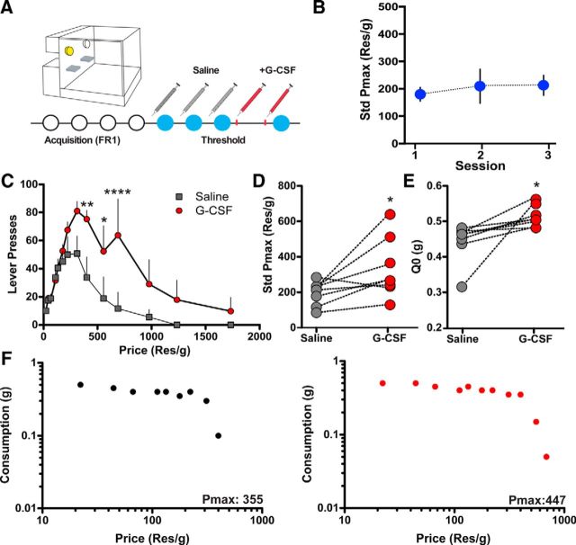
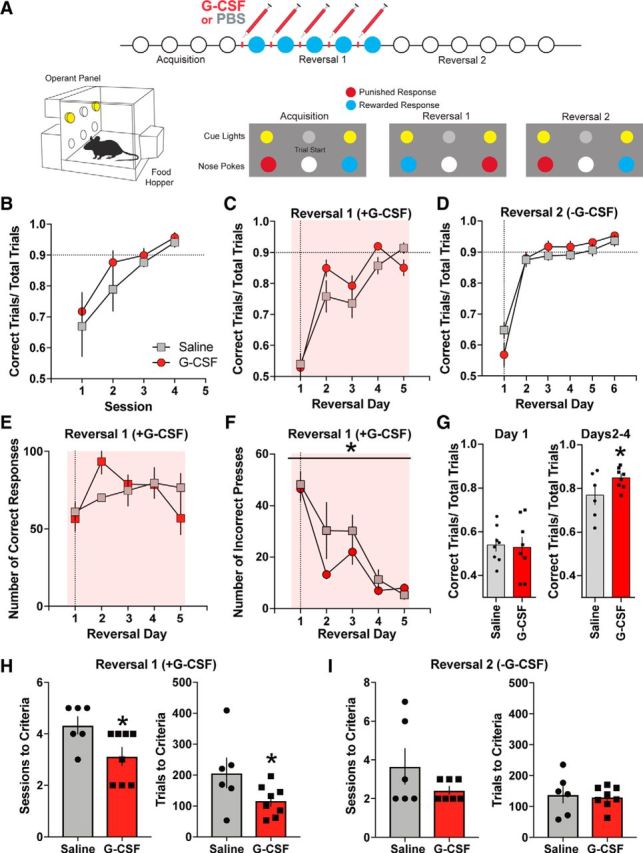
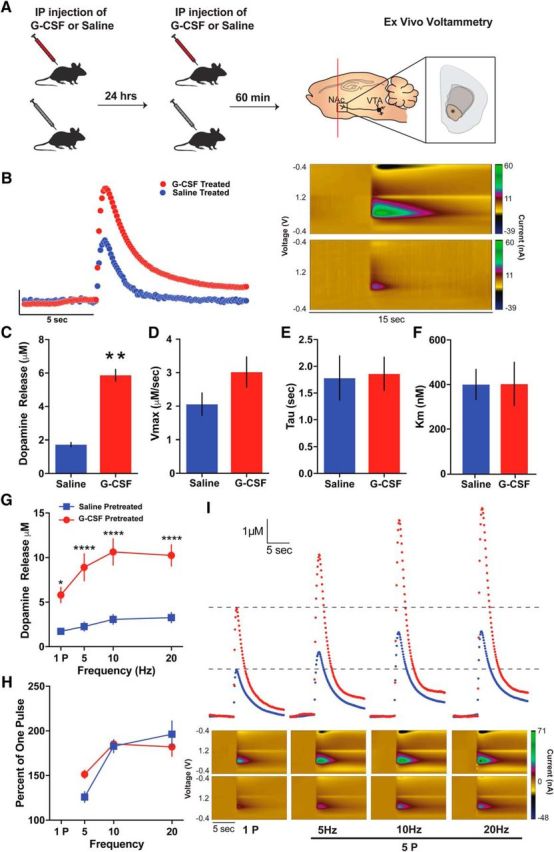
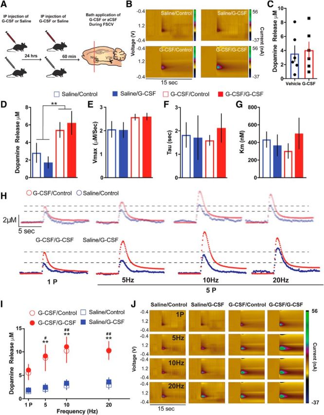
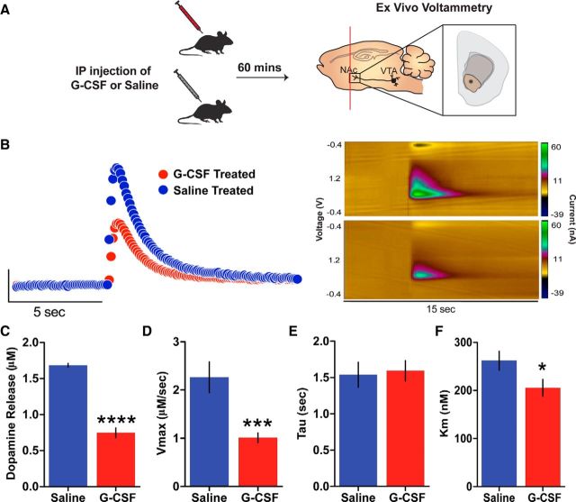
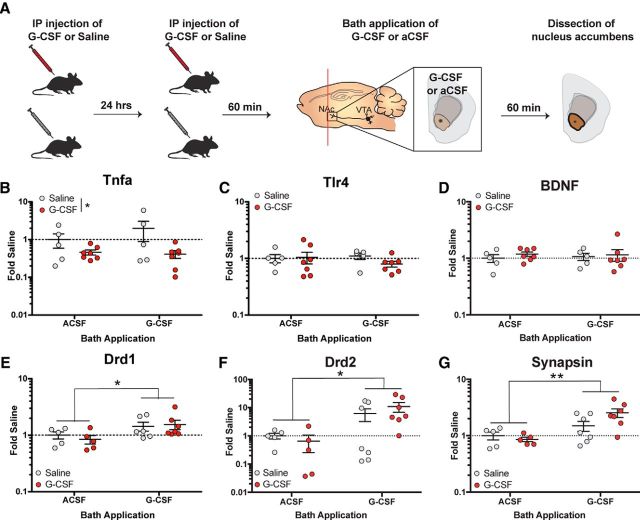
Deficits in motivation and cognition are hallmark symptoms of multiple psychiatric diseases. These symptoms are disruptive to quality of life and often do not improve with available medications. In recent years there has been increased interest in the role of the immune system in neuropsychiatric illness, but to date no immune-related treatment strategies have come to fruition. The cytokine granulocyte-colony stimulating factor (G-CSF) is known to have trophic and neuroprotective properties in the brain, and we recently identified it as a modulator of neuronal and behavioral plasticity. By combining operant tasks that assess discrete aspects of motivated behavior and decision-making in male mice and rats with subsecond dopamine monitoring via fast-scan cyclic voltammetry, we defined the role of G-CSF in these processes as well as the neural mechanism by which it modulates dopamine function to exert these effects. G-CSF enhanced motivation for sucrose as well as cognitive flexibility as measured by reversal learning. These behavioral outcomes were driven by mesolimbic dopamine system plasticity, as systemically administered G-CSF increased evoked dopamine release in the nucleus accumbens independent of clearance mechanisms. Importantly, sustained increases in G-CSF were required for these effects as acute exposure did not enhance behavioral outcomes and decreased dopamine release. These effects seem to be a result of the ability of G-CSF to alter local inflammatory signaling cascades, particularly tumor necrosis factor α. Together, these data show G-CSF as a potent modulator of the mesolimbic dopamine circuit and its ability to appropriately attend to salient stimuli.SIGNIFICANCE STATEMENT Emerging evidence has highlighted the importance of the immune system in psychiatric diseases states. However, the effects of peripheral cytokines on motivation and cognitive function are largely unknown. Here, we report that granulocyte-colony stimulating factor (G-CSF), a pleiotropic cytokine with known trophic and neuroprotective properties in the brain, acts directly on dopaminergic circuits to enhance their function. These changes in dopaminergic dynamics enhance reward learning and motivation for natural stimuli. Together, these results suggest that targeting immune factors may provide a new avenue for therapeutic intervention in the multiple psychiatric disorders that are characterized by motivational and cognitive deficits.

Keywords: cytokine; dopamine; immune system; learning and memory; motivation; voltammetry.


Figures

Figure 1.
Figure 1.
G-CSF increases the motivation for highly salient rewards. A, Timeline of behavioral economic assessment of sucrose reinforcement. Animals were injected with saline before behavioral testing for three sessions to establish a baseline. To determine the effects of G-CSF animals were injected with G-CSF twice, once 24 h before and a second time 60 min before, and the effect on economic parameters was determined (n = 8 per group). B, Pmax did not change across time in the saline treatment sessions. C, The number of lever presses over increasing price for sucrose. Animals treated with G-CSF emitted more lever presses overall than their pretreatment baseline. D, Standardized Pmax values highlighting that G-CSF within-subject increased the maximal price an animal would pay in effort to obtain sucrose pellets. E, G-CSF treatment also increased Q0, a measure of consumption at a minimally constraining price. These data highlight that G-CSF, a peripheral cytokine, is capable of enhancing the motivational properties of salient stimuli. F, Representative data from an animal treated with saline (left) and then G-CSF (right). G-CSF increased the Pmax for sucrose. *p < 0.05, **p < 0.01, ****p < 0.0001 from saline treated. All data are presented as mean SEM.
Figure 2.
Figure 2.
G-CSF enhances reversal learning. A, Timeline and experimental design. Animals were injected with either PBS or G-CSF before each Reversal 1, but not Reversal 2, test session (top). Representative lever-reward and lever-punishment contingencies (bottom; n = 6–8 per group). B, Number of correct trial to total number of trials ratio during acquisition. C, Reversal learning was enhanced by G-CSF treatment. D, Saline injections were given before each session during Reversal 2. Prior G-CSF treatment did not alter performance during Reversal 2. E, Total number of correct responses was not altered by G-CSF treatment. F, Number of incorrect responses was reduced following G-CSF administration in Reversal 1. G, G-CSF administration increased percentage correct response only after the second day of Reversal 1. H, Number of sessions (left) and number of trials to criteria were significantly decreased during Reversal 1 in the G-CSF-treated group. I, G-CSF administration did not affect number of sessions (left) and number of trials to criteria (right) during Reversal 2. *p < 0.05 from G-CSF. All data are presented as mean SEM.
Figure 3.
Figure 3.
G-CSF enhances presynaptic dopamine release in the nucleus accumbens. A, Timeline of G-CSF injections. Animals were injected with either saline or G-CSF 24 h and then 60 min before ex vivo voltammetry (left; n = 3–6 per group). FSCV recordings of subsecond dopamine release in the NAc (right). B, Current versus time plots (left) and color plots showing the presence of dopamine after single-pulse stimulation, as indicated by its oxidation at +0.6 V and reduction at −0.2 V (right). C, Group data showing enhanced dopamine release in the G-CSF-treated animals. D, There was no difference in maximal dopamine uptake (Vmax), (E) dopamine clearance (tau), or (F) the ability of dopamine to bind to the dopamine transporter (Km). G, Dopamine release evoked by multiple-pulse stimulations was increased in the G-CSF-treated animals. H, Dopamine release represented as a percentage of one pulse. I, Current versus time plots showing increased dopamine release to increasing stimulation frequency (top). Color plots showing dopamine oxidation/reduction in response to increasing stimulations (bottom). *p < 0.05, **p < 0.01, ***p < 0.001, ****p < 0.0001 from saline treated. All data are presented as mean SEM.
Figure 4.
Figure 4.
G-CSF effects on dopamine release are not through direct actions on dopamine terminals. A, Timeline of G-CSF injections. Animals were injected with either saline or G-CSF 24 h and then 60 min before ex vivo voltammetry (left; n = 3–6 per group). B, Color plots showing the presence of dopamine after a one-pulse stimulation (n = 3–6 per group). C, There were no effects of G-CSF bath application on dopamine release. D, Group data showing dopamine release following G-CSF or saline pretreatment and G-CSF or aCSF bath application. G-CSF pretreated groups showed elevated levels of NAc dopamine release. G-CSF bath application did not have a significant effect. **p < 0.01 from saline treated. E, G-CSF pretreatment or bath application did not alter maximal rates of dopamine uptake (Vmax), (F) dopamine clearance (tau), or (G) the ability of dopamine to bind to the dopamine transporter (Km). H, Current versus time plots showing increased dopamine release to increasing stimulation frequency. I, Dopamine release in response to multiple pulse stimulation parameters was increased in the G-CSF pretreated animals but not following G-CSF bath application. **p < 0.01 G-CSF/G-CSF versus Saline/Control, # p < 0.05 G-CSF/Control versus Saline/Control, ## p < 0.01 G-CSF/Control versus Saline/Control. J, Color plots showing the presence of dopamine following G-CSF pretreatment and bath application. All data are presented as mean SEM.
Figure 5.
Figure 5.
Acute G-CSF injection attenuates presynaptic dopamine release in the nucleus accumbens. A, Timeline of G-CSF or Saline injections. Animals were injected with either saline or G-CSF 60 min before ex vivo voltammetry (left; n = 3 per group). FSCV recordings of subsecond dopamine release in the NAc (right). B, Current versus time plots (left) and color plots showing the presence of dopamine after one-pulse (tonic) stimulation, as indicated by its oxidation at +0.6 V and reduction at −0.2 V (right). C, Group data showing reduced dopamine release in the acute G-CSF-treated animals. D, There was a significant decrease in maximal dopamine uptake (Vmax) and (E) the ability of dopamine to bind to the dopamine transporter (Km), but (F) no difference in dopamine clearance (tau) in the acute G-CSF-treated animals. *p < 0.05, **p < 0.01, ***p < 0.001, ****p < 0.0001 from saline treated. All data are presented as mean SEM.
Figure 6.
Figure 6.
G-CSF treatment reduces Tnfa expression and primes postsynaptic signaling. A, Timeline of G-CSF injections. Animals were injected with either saline or G-CSF 24 h and then 60 min (left) before G-CSF or aCSF bath application and dissection of NAc (right; n = 5–7 per group). B, NAc Tnfa mRNA levels calculated as fold-changes from the Saline/aCSF-treated controls. Tnfa expression in the NAc was decreased in the G-CSF pretreated group regardless of bath application. C,Tlr4 expression in the NAc was not affected by G-CSF pretreatment or bath application. D, Bdnf expression in the NAc was not altered by G-CSF. E, NAc Drd1 expression levels represented as fold-changes from saline/aCSF-treated controls. F, NAc Drd2 mRNA levels. G, Syn3 expression in the NAc. Expression levels of all three genes were elevated following G-CSF bath application regardless of pretreatment.*p < 0.05, **p < 0.01 between bath applications. All data are presented as mean SEM.

References

    1. Aberman JE, Salamone JD (1999) Nucleus accumbens dopamine depletions make rats more sensitive to high ratio requirements but do not impair primary food reinforcement. Neuroscience 92:545–552. 10.1016/S0306-4522(99)00004-4 - DOI - PubMed
    1. Austin MP, Mitchell P, Goodwin GM (2001) Cognitive deficits in depression: possible implications for functional neuropathology. Br J Psychiatry 178:200–206. 10.1192/bjp.178.3.200 - DOI - PubMed
    1. Basu S, Hodgson G, Katz M, Dunn AR (2002) Evaluation of role of G-CSF in the production, survival, and release of neutrophils from bone marrow into circulation. Blood 100:854–861. 10.1182/blood.V100.3.854 - DOI - PubMed
    1. Brennan FX, Beck KD, Servatius RJ (2004) Proinflammatory cytokines differentially affect leverpress avoidance acquisition in rats. Behav Brain Res 153:351–355. 10.1016/j.bbr.2003.12.025 - DOI - PubMed
    1. Calipari ES, Ferris MJ, Melchior JR, Bermejo K, Salahpour A, Roberts DC, Jones SR (2014) Methylphenidate and cocaine self-administration produce distinct dopamine terminal alterations. Addict Biol 19:145–155. 10.1111/j.1369-1600.2012.00456.x - DOI - PMC - PubMed

Publication types

MeSH terms

LinkOut - more resources