. 2018 Sep 11;115(37):E8717-E8726.
doi: 10.1073/pnas.1807105115. Epub 2018 Aug 27.

Mechanisms of enhanced drug delivery in brain metastases with focused ultrasound-induced blood-tumor barrier disruption

Affiliations

Mechanisms of enhanced drug delivery in brain metastases with focused ultrasound-induced blood-tumor barrier disruption

Costas D Arvanitis et al. Proc Natl Acad Sci U S A. .

Abstract

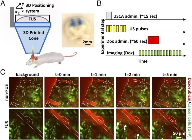
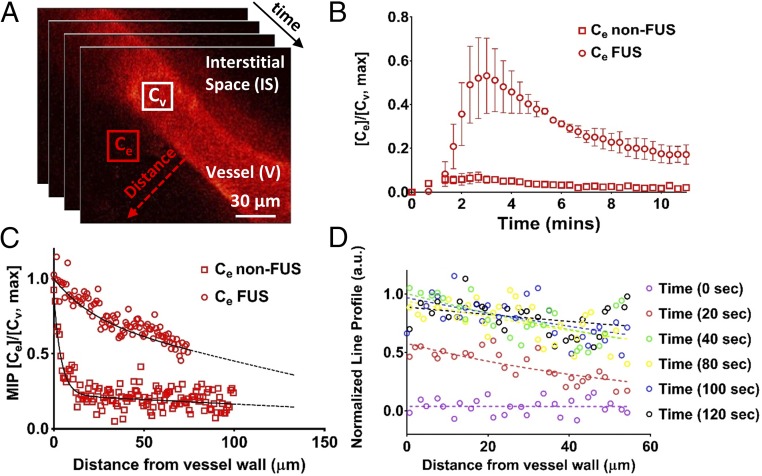
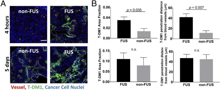
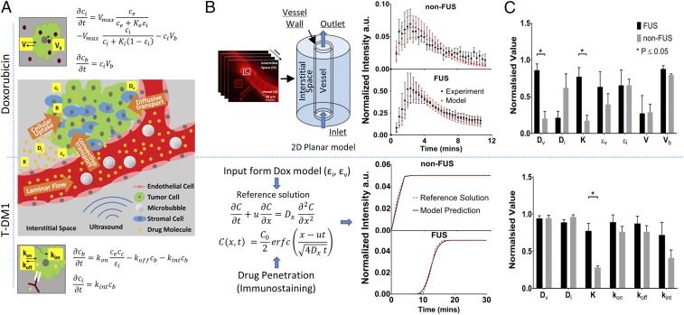
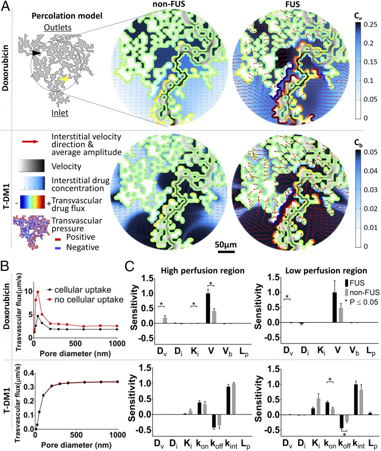
Blood-brain/blood-tumor barriers (BBB and BTB) and interstitial transport may constitute major obstacles to the transport of therapeutics in brain tumors. In this study, we examined the impact of focused ultrasound (FUS) in combination with microbubbles on the transport of two relevant chemotherapy-based anticancer agents in breast cancer brain metastases at cellular resolution: doxorubicin, a nontargeted chemotherapeutic, and ado-trastuzumab emtansine (T-DM1), an antibody-drug conjugate. Using an orthotopic xenograft model of HER2-positive breast cancer brain metastasis and quantitative microscopy, we demonstrate significant increases in the extravasation of both agents (sevenfold and twofold for doxorubicin and T-DM1, respectively), and we provide evidence of increased drug penetration (>100 vs. <20 µm and 42 ± 7 vs. 12 ± 4 µm for doxorubicin and T-DM1, respectively) after the application of FUS compared with control (non-FUS). Integration of experimental data with physiologically based pharmacokinetic (PBPK) modeling of drug transport reveals that FUS in combination with microbubbles alleviates vascular barriers and enhances interstitial convective transport via an increase in hydraulic conductivity. Experimental data demonstrate that FUS in combination with microbubbles enhances significantly the endothelial cell uptake of the small chemotherapeutic agent. Quantification with PBPK modeling reveals an increase in transmembrane transport by more than two orders of magnitude. PBPK modeling indicates a selective increase in transvascular transport of doxorubicin through small vessel wall pores with a narrow range of sizes (diameter, 10-50 nm). Our work provides a quantitative framework for the optimization of FUS-drug combinations to maximize intratumoral drug delivery and facilitate the development of strategies to treat brain metastases.

Keywords: blood–brain/blood–tumor barrier; brain tumor; drug transport; focused ultrasound; pharmacokinetics.


Conflict of interest statement

Conflict of interest statement: R.K.J. received an honorarium from Amgen and consultant fees from Merck, Ophthotech, Pfizer, SPARC, SynDevRx, and XTuit; owns equity in Enlight, Ophthotech, SynDevRx, and XTuit; served on the Board of Directors of XTuit; and serves on the Boards of Trustees of Tekla Healthcare Investors, Tekla Life Sciences Investors, Tekla Healthcare Opportunities Fund, and Tekla World Healthcare Fund. Neither any reagent nor any funding from these organizations was used in this study.

Figures

Fig. 1.
Fig. 1.
FUS enhances doxorubicin extravasation in BT474-Gluc brain tumors. (A) FUS system and experimental setup. (Inset) Image of trypan blue extravasation in gross pathology of a coronal plane section after FUS-BTB disruption in healthy mice (480-kPa peak negative pressure). (B) Schematic illustration of the drug administration protocol. Ultrasound contrast agent (USCA–Definity; Lantheus Medical Imaging) was administered as a bolus. (C) Representative sequential images from intravital multiphoton microscopy of doxorubicin distribution in the breast cancer BM model with (Lower) and without (Upper) FUS-BTB disruption. Red, doxorubicin autofluorescence; green, GFP-positive BT474-Gluc cancer cells.
Fig. 2.
Fig. 2.
FUS enhances doxorubicin (Dox) penetration and promotes convective transport in BT474-Gluc brain tumors. (A) Sequential intravital multiphoton microscopy of Dox autofluorescence. Approximately 50 images were acquired at 20-s intervals during i.v. injection of 150 µL of Dox at a concentration of 7 mg/mL over 30 s (7.5 mg/kg). Three images were acquired before the Dox administration to establish background fluorescence. (B) Temporal evaluation of Dox extravasation with and without FUS-BTB disruption. Cv and Ce are the mean pixel intensity of the vessel and the extravascular space, respectively. The center of the region of interest (20 × 20 pixels) was 20 µm from the vessel wall. The plots show means ± SEM (n = 4 for each condition, i.e., non-FUS and FUS). The maximum mean fluorescence for the FUS and non-FUS was 0.52 ± 0.15 and 0.07 ± 0.02, a sevenfold difference. (C) Dox penetration from a line profile perpendicular to vessel wall (red dotted arrow in A). The plot shows the normalized maximum intensity projection (MIP) across the series of images. The dotted line shows a regression fitted to the data from four different animals for each condition (non-FUS and FUS). (D) Representative data of the temporal evolution of the normalized intensity of the line profile (red dotted arrow in A). For consistency in the notation of the experiments/modeling, Cv is the Dox intensity/concentration in the vessel, Ce is the Dox intensity/concentration in the extracellular/interstitial space.
Fig. 3.
Fig. 3.
FUS-BTB disruption increases early extravasation and penetration of T-DM1 in BT474-Gluc brain tumors. (A) Representative microscopy data of T-DM1 extravasation with and without FUS at 4 h and 5 d. (B) Quantification of the T-DM1 extravasation (Left) and penetration (Right) with and without FUS at 4 h (Upper) and 5 d (Lower) posttreatment. The plots show means ± SEM (n = 6). (Scale bar, 100 µm.) Parametric Student’s t test for P < 0.05 (Prism 6; GraphPad). n.s., not significant.
Fig. 4.
Fig. 4.
Quantification of transvascular transport via mathematical modeling demonstrates multifold increase in effective diffusion coefficient (4.3-fold) and in hydraulic conductivity (4.5-fold) after FUS-BTB disruption. Vascular perfusion and cellular transport dictate interstitial drug transport after FUS-BTB disruption in BT474-Gluc brain tumors. (A) Schematic illustrating the transport of the anticancer agents from the vessel to the interstitial space along with the studied model parameters and agent-specific cellular uptake model equations. (Upper) Convection–diffusion–reaction model following Michaelis–Menten kinetics with binding of doxorubicin to DNA (Vb). (Lower) Convection–diffusion–reaction model for T-DM1. Excellent fit was observed for both doxorubicin and T-DM1. (B, Upper) The time dependence of doxorubicin extravasation from the fitted and experimental data for non-FUS and FUS-BBB/BTB disruption groups. (B, Lower) Parameter fit methodology for T-DM1 and fitted data from two different experiments. The fitted vascular and interstitial effective porosity (fraction of surface area occupied by pores) from the doxorubicin model was used as input to the T-DM1 fitting (i.e., same animal model). (C) Normalized parameter fit for non-FUS and FUS-BBB/BTB disruption groups (Upper, doxorubicin; Lower, T-DM1). The values for each parameter were normalized to maximum to be displayed on the same plot. The exact numbers and their units are shown in Tables 1 and 2 for doxorubicin and T-DM1, respectively. The plots show means ± SEM from fitted values from four different experiments for each condition.
Fig. 5.
Fig. 5.
Structurally heterogeneous mathematical tumor model predicts that FUS-BTB disruption overcomes transvascular transport barriers in brain tumor microenvironment and reveals the critical role of cancer cell transmembrane transport for effective uptake. (A) Modeling of vascular heterogenous perfusion using percolation model. Interstitial velocity, transvascular pressure difference (thresholded to highlight positive and negative regions), drug transvascular flux, and interstitial drug concentration captured at the time where drug reached its peak intensity [5 min for doxorubicin (Dox)—bolus administration—and 20 min for T-DM1]. The pore diameter used was 50 nm. (B) Transvascular flux for the Dox and T-DM1 (with FUS) with and without the cellular uptake. (C) Sensitivity analysis of the model parameters. The plots show means ± SEM from fitted values from four different experiments for each condition. The yellow arrow shows high perfusion region; the black arrow shows low perfusion region.
Fig. 6.
Fig. 6.
FUS-BTB disruption increases doxorubicin transmembrane transport in endothelial and extravascular cells. (A) Representative doxorubicin images at 0.3 and 5 min after doxorubicin administration showing vessels, interstitial space (IS), and segmented endothelial (EC) and extravascular cells (EVC). (B) Quantification of intracellular doxorubicin kinetics for endothelial and interstitial cells. Cv is the doxorubicin intensity/concentration in the vessel, and Ci is the intracellular doxorubicin intensity/concentration. The plots show means ± SEM (n = 6) from each cell population. (C) Fitted rate of endothelial cellular transmembrane transport and drug bound to DNA.

References

    1. Kodack DP, Askoxylakis V, Ferraro GB, Fukumura D, Jain RK. Emerging strategies for treating brain metastases from breast cancer. Cancer Cell. 2015;27:163–175. - PMC - PubMed
    1. Lin X, DeAngelis LM. Treatment of brain metastases. J Clin Oncol. 2015;33:3475–3484. - PMC - PubMed
    1. Sperduto PW, et al. Summary report on the graded prognostic assessment: An accurate and facile diagnosis-specific tool to estimate survival for patients with brain metastases. J Clin Oncol. 2012;30:419–425. - PMC - PubMed
    1. Kabraji S, et al. Drug resistance in HER2-positive breast cancer brain metastases: Blame the barrier or the brain? Clin Cancer Res. 2018;24:1795–1804. - PMC - PubMed
    1. Lockman PR, et al. Heterogeneous blood-tumor barrier permeability determines drug efficacy in experimental brain metastases of breast cancer. Clin Cancer Res. 2010;16:5664–5678. - PMC - PubMed

Publication types

MeSH terms