. 2018 Sep 11;115(37):E8668-E8677.
doi: 10.1073/pnas.1810498115. Epub 2018 Aug 27.

BRAF/MAPK and GSK3 signaling converges to control MITF nuclear export

Affiliations

BRAF/MAPK and GSK3 signaling converges to control MITF nuclear export

Kao Chin Ngeow et al. Proc Natl Acad Sci U S A. .

Abstract

The close integration of the MAPK, PI3K, and WNT signaling pathways underpins much of development and is deregulated in cancer. In principle, combinatorial posttranslational modification of key lineage-specific transcription factors would be an effective means to integrate critical signaling events. Understanding how this might be achieved is central to deciphering the impact of microenvironmental cues in development and disease. The microphthalmia-associated transcription factor MITF plays a crucial role in the development of melanocytes, the retinal pigment epithelium, osteoclasts, and mast cells and acts as a lineage survival oncogene in melanoma. MITF coordinates survival, differentiation, cell-cycle progression, cell migration, metabolism, and lysosome biogenesis. However, how the activity of this key transcription factor is controlled remains poorly understood. Here, we show that GSK3, downstream from both the PI3K and Wnt pathways, and BRAF/MAPK signaling converges to control MITF nuclear export. Phosphorylation of the melanocyte MITF-M isoform in response to BRAF/MAPK signaling primes for phosphorylation by GSK3, a kinase inhibited by both PI3K and Wnt signaling. Dual phosphorylation, but not monophosphorylation, then promotes MITF nuclear export by activating a previously unrecognized hydrophobic export signal. Nonmelanocyte MITF isoforms exhibit poor regulation by MAPK signaling, but instead their export is controlled by mTOR. We uncover here an unanticipated mode of MITF regulation that integrates the output of key developmental and cancer-associated signaling pathways to gate MITF flux through the import-export cycle. The results have significant implications for our understanding of melanoma progression and stem cell renewal.

Keywords: GSK3; MAPK; MITF; melanoma; nuclear export.


Conflict of interest statement

The authors declare no conflict of interest.

Figures
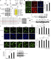
Fig. 1.
Fig. 1.
GSK3 phosphorylates MITF S69 to control MITF subcellular localization. (A) MITF posttranslational modifications. (B) Immunofluorescence of SKmel28 cells following treatment with 1 μM BIO or DMSO for 18 h. Cells were stained with DAPI (blue) and antibodies against MITF (green) or β-catenin (red). Quantification (Right). n > 70 per condition. Error bars represent SEM. Two-tailed t test: ****P < 0.0001. (C) Western blot of SKmel28 cells following treatment with 1 μM BIO, 1 μM of SB-675259-M, or DMSO as in A. (D) Amino acid sequence alignment of MITF. In the GSK3 consensus, X = any amino acid. (E, Left) Twenty-one residue peptides corresponding to MITF amino acids 61 to 81 used for the SPOT kinase assay immobilized on a cellulose support membrane. (E, Right) SPOT kinase assay using purified GSK3β. A map showing the identity of the 10 tested peptides (Left) corresponding to peptide spots on the membrane imaged in UV light (Middle), and the kinase assay (Right). (F) Western blot of 501mel cells ectopically expressing indicated MITF-FLAG WT and mutants. (G) Western blot of extracts from 501mel cells ectopically expressing indicated MITF-FLAG WT and mutants treated or not with calf intestinal phosphatase (CIAP). (H and I) Fluorescence images of 501mel cells ectopically expressing GFP-MITF WT and indicated mutants (green). (Scale bars: 10 μm.) Quantification (Right). n > 40 per condition. Error bars represent SEM. Two-tailed t test (Upper) and one-way ANOVA with post hoc Tukey test: ****P < 0.0001, NS, not significant, P > 0.05. (J) Luciferase assay with MITF-FLAG WT and mutants and a TYR promoter-Luc reporter cotransfected into HeLa cells. Error bars represent SEM. Two-tailed t test: ****P < 0.0001. Western blot shows relative expression of WT and mutant MITF-FLAG proteins.
Fig. 2.
Fig. 2.
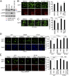
BRAF/MAPK signaling redirects MITF to the cytoplasm. (A) Western blot or (B) immunofluorescence of 501mel cells following 200 nM TPA treatment (1 h) in the presence or absence of 10 μM U0126 (3 h). After fixation and permeabilization, the cells were stained with the nucleic acid stain DAPI (blue) and antibodies against MITF (green) or phospho-ERK (red). n >100 for each condition. (C) Immunofluorescence of 501mel cells treated with 200 nM TPA (1 h) and/or 1 μM BIO. n > 40 cells per condition. (D) Fluorescence assay of 501mel cells ectopically expressing WT or mutant MITF-GFP and treated with 200 nM TPA for 1 h as indicated. n > 49 for each condition. (E) Immunofluorescence of Flip-in HeLa cells engineered to express doxycycline-inducible BRAFV600E-FLAG ectopically expressing GFP-MITF WT or the S73A mutant (green). Twenty-four hours posttransfection, cells were treated with 2 ng/mL doxycycline for 24 h and treated as indicated with 10 μM U0126 for 3 h. Anti-FLAG-BRAF (red). n > 10 cells per condition. Quantification by two-tailed t test or one-way ANOVA with post hoc Tukey test (E):*P < 0.05; **P < 0.01; ***P < 0.001; ****P < 0.0001; NS, not significant, P > 0.05; error bars represent SEM.
Fig. 3.
Fig. 3.
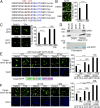
MITF has a nuclear export signal. (A) Alignment of MITF sequences in the vicinity of S69 and S73 from different species. Hydrophobic residues highlighted in blue indicate the putative NES. (B) Immunofluorescence of endogenous MITF in 501mel cells treated with 20 nM LMB for 3 h as indicated. n > 80 cells per condition. (C) Fluorescence images of 501mel cells ectopically expressing WT GFP-MITF (green) treated with 200 nM TPA for 1 h and/or 20 nM LMB for 3 h. n > 49 per condition. (D) CRM1 pull-down assay using bacterially expressed HIS-Tagged MITF (1–105) bound to Ni-NTA beads. All proteins were bacterially expressed and purified, and, after pull-down, CRM1 and RAN were detected by Western blotting using specific antibodies. Purified MITF was visualized by Coomassie staining (Lower). (E and F) Fluorescence assay of 501mel cells transfected with indicated GFP reporters. n > 43 per condition. Quantification by two-tailed t test: **P < 0.01; ****P < 0.0001; NS, not significant, P > 0.05; error bars represent SEM.
Fig. 4.
Fig. 4.
Regulated nuclear export of MITF-A isoforms. (A) Immunofluorescence using anti-MITF antibody of HeLa cells transfected with expression vectors for MITF-A isoform treated with indicated drugs (20 nM LMB; 10 μM U0126; 200 nM Torin 1; 1 μM BIO) or expressing the L80A mutant. n > 20 cells per condition. (B) Immunofluorescence using anti-MITF of ARP19 (RPE) cells with or without 20 nM LMB. n > 20 cells per condition. (Scale bars: 10 μm.) Quantifications by two-tailed t test: **P < 0.01; ***P < 0.001; ****P < 0.0001. Error bars represent SEM.
Fig. 5.
Fig. 5.
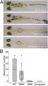
MITF S69 is required for melanocyte development. (A) Representative images of mitfa-null nacre embryos injected with plasmids encoding Mitfa WT or indicated mutants taken 5 d postfertilization (dpf). (B) Quantification of the number of rescued melanocytes per embryo. Mitfa WT (number of embryos n = 11, mean melanocytes per embryo = 9.9); S69A mutant (number of embryos = 29, mean number of melanocytes per embryo = 6.1); S69E number of embryos = 25, mean = 0). For WT vs. S69A: no significant difference by one-way ANOVA test [95% CI: (−0.876, 8.418)] (experimental replicates n = 3).
Fig. 6.
Fig. 6.
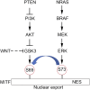
Model illustrating potential regulation of MITF nuclear export.

Comment in

References

    1. Garraway LA, et al. Integrative genomic analyses identify MITF as a lineage survival oncogene amplified in malignant melanoma. Nature. 2005;436:117–122. - PubMed
    1. Lister JA, et al. A conditional zebrafish MITF mutation reveals MITF levels are critical for melanoma promotion vs. regression in vivo. J Invest Dermatol. 2014;134:133–140. - PMC - PubMed
    1. Hodgkinson CA, et al. Mutations at the mouse microphthalmia locus are associated with defects in a gene encoding a novel basic-helix-loop-helix-zipper protein. Cell. 1993;74:395–404. - PubMed
    1. Widlund HR, et al. Beta-catenin-induced melanoma growth requires the downstream target microphthalmia-associated transcription factor. J Cell Biol. 2002;158:1079–1087. - PMC - PubMed
    1. Carreira S, et al. Mitf cooperates with Rb1 and activates p21Cip1 expression to regulate cell cycle progression. Nature. 2005;433:764–769. - PubMed

Publication types

Substances