. 2018 Aug 27;8(1):12870.
doi: 10.1038/s41598-018-30316-w.

Rapid Regeneration and Reuse of Silica Columns from PCR Purification and Gel Extraction Kits

Affiliations

Rapid Regeneration and Reuse of Silica Columns from PCR Purification and Gel Extraction Kits

Ying Zhou et al. Sci Rep. .

Abstract

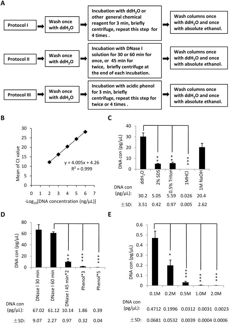
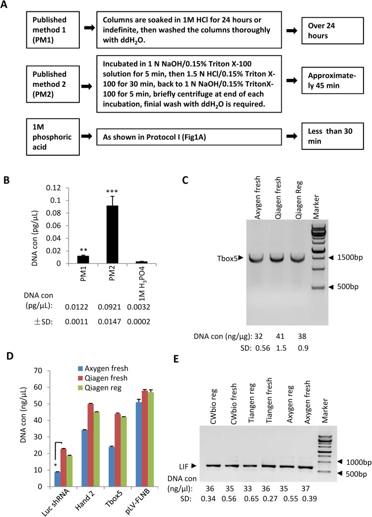
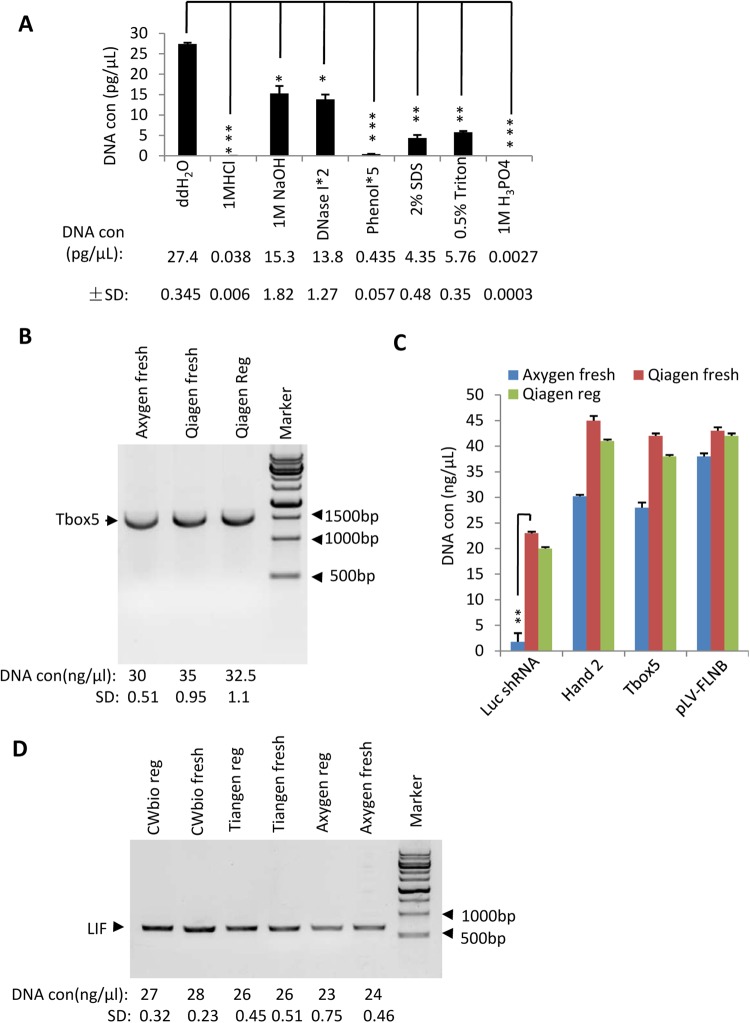
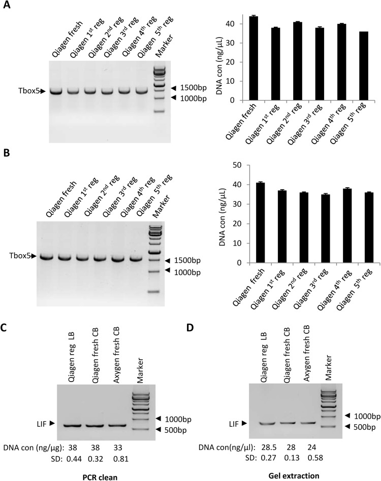
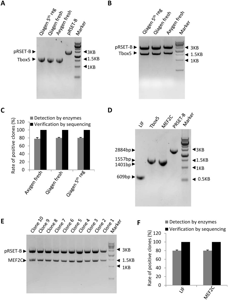
Silica columns from PCR purification and gel extraction kits are widely used in laboratories worldwide to assist in gene cloning. However, the use of these columns can generate plastic waste that has an environmental impact due to their one-off design and massive consumption. Thus, it is important to develop a novel method that can reduce the utilization of silica columns but not affect research efficiency. In this study, various chemical and nonchemical reagents were used to eliminate residual DNA within used columns from PCR purification and gel extraction kits. We show that phosphoric acid is the most effective reagent among those tested to remove DNA contamination from used columns. Columns regenerated using 1 M phosphoric acid have a DNA purification capability that is comparable to that of fresh columns. We demonstrate that silica columns can be regenerated and reused a minimum of five times. The lab-made buffers are compatible with the regenerated columns for DNA purification, and DNA that is prepared with the regenerated columns can be used for gene cloning without affecting the gene cloning efficiency. Thus, the use of this novel method greatly reduces the production of laboratory waste and benefits numerous laboratories worldwide.


Conflict of interest statement

The authors declare no competing interests.

Figures

Figure 1
Figure 1
Phosphoric acid is the most effective reagent for eliminating residual DNA from used Qiagen PCR purification kit columns. (A) A scheme showing the workflow for different protocols used in this study. (B) The standard curve that was plotted with the Ct values against –Log10 DNA concentration (ng/μL). Ten microliters of LIF DNA template at a concentration of 100 ng/μL was consecutively diluted at 1/10 for 8 times, and the diluted samples (starting from 1 * 10−2 ng/μL) were used for qPCR. Ct: Threshold cycles in quantitative PCR. (C) qPCR analysis to assess the amount of residual DNA eluted from the columns that were cleaned with different chemical reagents. LIF-contaminated columns were cleaned with indicated reagent based on protocol I. One microlitre of eluate was used to assess the presence of residual DNA by qPCR, with each sample assayed in triplicate. The obtained Ct values were converted into DNA concentration using the standard curve in B and were subjected to statistical analysis. The value of each column within the graph is presented in the bottom of the panel to clearly show the differences between the samples. (D) qPCR results for the amount of residual DNA from the columns that were cleaned with DNase I or acidic phenol. Contaminated columns were cleaned based on protocol II or III. qPCR assays were performed as in C. (E) qPCR showing DNA elimination for the used columns cleaned with different concentrations of phosphoric acid. Contaminated columns were cleaned with different concentrations of phosphoric acid as detailed in protocol I. qPCR assays performed as in C. Each column in C–E represents the mean ± SD of three independent experiments. *p < 0.05; **p < 0.01 and ***p < 0.001, where p was obtained by a one way ANOVA.
Figure 2
Figure 2
Regenerated PCR purification kit columns have a comparable capacity for DNA purification as fresh columns. (A) A scheme showing the workflow and duration for the published regeneration methods and for the 1 M phosphoric acid method. PM: published method. (B) Comparison of DNA elimination using 1 M phosphoric acid and the published methods. The used columns were cleaned with the protocol as shown in A. Thirty microlitres of eluate was obtained from the regenerated columns, 1 μL of which was used for qPCR. The data were processed as shown in Fig. 1C. (C) Comparison of DNA purification efficacy between the regenerated and fresh columns. A 50 μL Tbox 5 PCR reaction was purified with the regenerated or fresh columns, and the DNA concentrations were measured with a Nanophotometer (Thermo Scientific) and then subjected to a statistical analysis. The DNA quality was verified by agarose gel electrophoresis and imaged using a Bio-Rad ChemiDoc XRS + System. Data acquisition and settings are described as in the materials and methods section. The full-length image is presented in Supplementary Fig. S2. The DNA concentration for each sample is shown beneath the gel picture. (D) The regenerated columns showed a similar capacity for purifying different sizes of DNA as the fresh columns. DNA from a 50 μL PCR reaction was purified and quantified as in C. Luc shRNA, Luciferase shRNA cDNA, 70 bp; Hand2, 654 bp, Tbox 5, 1557 bp; pLV-FLNB, 17360 bp. (E) Comparison of DNA purification efficacy between the regenerated and fresh columns from different PCR purification kits. DNA from a 50 μL LIF PCR reaction was purified using regenerated or fresh columns, and the data were processed and presented as in C. Each column in B and D represents the mean ± SD of three independent experiments. *p < 0.05; **p < 0.01 and ***p < 0.001, where p was obtained by a one way ANOVA.
Figure 3
Figure 3
Analysis of DNA extraction efficacy using regenerated columns from gel extraction kits. (A) qPCR analysis for the residual DNA from Qiagen columns regenerated using different reagents. LIF-contaminated columns were cleaned with the indicated reagents based on the protocols in Fig. 1A. Thirty microlitres of eluate was obtained from the regenerated columns, 1 μL of which was used for qPCR. The data were processed and presented as in Fig. 1C. (B) Comparison of the DNA extraction efficacy between the regenerated and fresh columns from gel extraction kits. A 50 μL PCR reaction was used for agarose gel electrophoresis, and DNA extraction was performed using the regenerated or fresh columns according to the gel extraction kit manual. The extracted DNA was quantified with a Nanophotometer and verified by agarose gel electrophoresis as shown in Fig. 2C. The full-length image is presented in Supplementary Fig. S3. (C) The regenerated columns showed a comparable ability to purify different sizes of DNA as the fresh columns. A 50 μL PCR reaction was used for agarose gel electrophoresis, which was purified and measured as in (B). (D) Comparison of DNA purification efficacy between the regenerated and fresh columns from different commercial kits. A 50 μL LIF PCR reaction was used for agarose gel electrophoresis, and the DNA was extracted from the gel with regenerated or fresh columns from different gel extraction kits. The data were processed and presented as in B. Each column in A and C represents the mean ± SD of three independent experiments. *p < 0.05; **p < 0.01 and ***p < 0.001, where p was obtained by a one way ANOVA.
Figure 4
Figure 4
Disposable columns from Qiagen PCR purification and gel extraction kits can be repeatedly regenerated and reused a minimum of five times. (A) DNA purification efficacy for the repeatedly regenerated PCR purification kit columns. DNA-contaminated columns were cleaned with 1 M phosphoric acid and used for PCR product (TBox 5) purification; the same columns were regenerated and reused four additional times. The concentration of obtained DNA for each cycle was measured with a Nanophotometer and the data are presented as gel pictures (left) and in a graph (right). (B) The analysis of DNA extraction efficacy for the repeatedly regenerated gel extraction kit columns. DNA-contaminated columns were regenerated as in A, where a 50 μL PCR reaction was used for agarose gel electrophoresis. DNA extraction from agarose gel was performed according to the gel extraction kit manual. The data were processed as in A. The full length images for A (left) and B (left) are presented in Supplementary Fig. S4A,B. (C) Comparison of DNA purification efficacy between lab-made and commercial buffers. The lab-made buffers (LB) were used for DNA purification for regenerated Qiagen columns, whereas the commercial buffers (CB) were used for DNA purification for the fresh Qiagen or Axygen kit columns. The assays were performed as shown in Fig. 2C. (D) Comparison of DNA extraction efficacy between lab-made and commercial buffers. The lab-made buffers (LB) were used for DNA extraction from agarose gel using regenerated Qiagen columns, whereas the commercial buffers (CB) were used for DNA extraction from agarose gel using fresh Qiagen or Axygen kit columns. The assays were performed as shown in Fig. 3B. The protocol for DNA purification with lab-made buffers is described in the materials and methods section. Each column in A (right) and B (right) represents the mean ± SD of three independent experiments.
Figure 5
Figure 5
DNA prepared with the regenerated columns does not affect cloning efficiency. (A) Agarose gel electrophoresis image for the DNA samples purified with the regenerated and fresh columns. One microgram of DNA was digested with BamHI and HindIII, followed by agarose gel electrophoresis and DNA extraction. (B) Electrophoresis image for the positive clones digested with BamHI and HindIII. The DNA purified with the fresh or regenerated columns was used for gene cloning. Fifty colonies were picked from LB plates for plasmid preparation. Positive clones were detected by digestion with BamHI and HindIII, and one positive clone for each sample was loaded into an agarose gel for electrophoresis. (C) The preparation of DNA using regenerated columns does not affect cloning efficiency. The number of positive clones obtained in B was used for statistical analysis; the positive clones were verified by DNA sequencing. (D) Electrophoresis image for the DNA samples purified with the regenerated columns. Tbox 5 DNA digested with BamHI and HindIII was purified from agarose gels with the regenerated columns. The Tbox 5-contaminated columns were further regenerated with 1 M phosphoric acid and used to purify the MEF2C gene, which was also digested with BamHI and HindIII. The obtained DNA was detected by agarose gel electrophoresis. (E) Electrophoresis image for MEF2C-positive clones digested with BamHI and HindIII. Positive clones were screened as in B, and 10 positive clones digested with restriction enzymes were loaded into an agarose gel for electrophoresis. (F) The small amount of contaminating DNA has no chance of be cloned into the plasmid. The number of positive clones was analysed as in C. The full-length images for A, B, D and E are presented in Supplementary Fig. S5A, B, D and E. Each column in C and F within the graph represents the mean ± SD of three independent experiments.

Similar articles

Cited by

References

    1. Jackson D, Symons R, Berg P. Biochemical method for inserting new genetic information into DNA of Simian Virus 40: Circular SV40 DNA molecules containing lambda phage genes and the galactose operon of Escherichia coli. Proc. Natl. Acad. Sci. USA. 1972;69:2904–2909. doi: 10.1073/pnas.69.10.2904. - DOI - PMC - PubMed
    1. Lobban P, Kaiser A. Enzymatic end-to end joining of DNA molecules. J. Mol. Biol. 1973;78:453–471. doi: 10.1016/0022-2836(73)90468-3. - DOI - PubMed
    1. Cohen S, Chang A, Boyer H, Helling R. Construction of biologically functional bacterial plasmids in vitro. Proc. Natl. Acad. Sci. USA. 1973;70:3240–3244. doi: 10.1073/pnas.70.11.3240. - DOI - PMC - PubMed
    1. Nathans D, Smith HO. Restriction endonucleases in the analysis and restructuring of DNA molecules. Ann. Rev. Biochem. 1975;44:273–293. doi: 10.1146/annurev.bi.44.070175.001421. - DOI - PubMed
    1. Johnson IS. Human insulin from recombinant DNA technology. Science. 1983;219:632–637. doi: 10.1126/science.6337396. - DOI - PubMed

Publication types

LinkOut - more resources