Case Reports
. 2018;22(2):118-123.
doi: 10.5114/wo.2018.76235. Epub 2018 Jun 13.

Giant cranionasal epithelioid haemangioendothelioma with invasive growth pattern mimicking a skull base chondrosarcoma

Affiliations
Case Reports

Giant cranionasal epithelioid haemangioendothelioma with invasive growth pattern mimicking a skull base chondrosarcoma

Kelvin Piña Batista et al. Contemp Oncol (Pozn). 2018.

Abstract

Epithelioid haemangioendothelioma (EHE) is a rare low-grade vascular neoplasm that is composed of mostly epithelioid cells. EHE may arise as a solitary tumour or in the form of multiple body lesions, and commonly occurs in soft tissues, liver, pleura, lung, peritoneum, lymph nodes, breast, and many other sites. EHE in the cranionasal region is extremely rare. There are very few reports of cases of skull-base EHE. We discuss an extremely rare presentation of an aggressive EHE that originated from the sellar region. Based on literature review, our patient is the first reported case of a giant solitary EHE with prepontine cistern invasion and abducens nerve encroachment mimicking a chondrosarcoma. We treated this rare tumour by near subtotal surgical excision with subsequent radiotherapy, considering that complete tumour resection with free margins in both cavernous sinus and clival region avoiding neural and vascular structure encroachment becomes technically difficult.

Keywords: cranionasal; epithelioid haemangioendothelioma; skull base; vascular tumour.


Conflict of interest statement

The authors declare no conflict of interest.

Figures
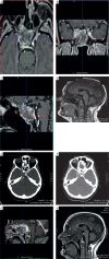
Fig. 1
Fig. 1
Preoperative computed tomography and MR images of the tumour. A–B) Pre-contrast and post-contrast computed tomography. Yellow arrow shows posterior clival and prepontine cistern invasion. C–H) MRI using T1-weighted imaging (T1WI) was hyperintense with heterogeneous isointensity to grey matter showing an expansile lesion with intracranial and extracranial components, its epicenter being in the sphenoid sinus and right cavernous sinus (red asterisk). The mass measured 6.5 × 4.2 × 3.7 cm with erosion of the optic canal (blue arrow), middle cranial fossa, clivus, and petro-occipital fissure and with protrusion to the sphenoid sinus and nasopharynx (white arrow). Both the pituitary gland and the chiasm were displaced superiorly. The mass also caused effacement of the prepontine cistern with encroachment on the right internal carotid artery (ICA) and Dorello’s canal, while T2WI showed a high hyperintensity along with partial isointensity to the grey matter, which suggested the presence of haemorrhage
Fig. 2
Fig. 2
Histopathological findings. A–B) Immunohistochemical staining showing positivity of the spindle to epithelioid tumour cells embedded in myxofibro-hyaline stroma (400× and 200×, respectively), demonstrating intracytoplasmic lumina (yellow arrow) with erythrocytes (black arrow). C, D, E, and F) Tumour cells were positive for CD34, CD31, Factor VIII, and CD99, respectively (400×). G) Ki67 proliferation index was 10% (400×). H) The immunohistochemical stains revealed negativity for cytokeratin (400×)
Fig. 3
Fig. 3
Postoperative MRI showing minimal residual tumour (white arrow)

References

    1. Fernandes AL, Ratilal B, Mafra M, Magalhaes C. Aggressive intracranial and extra-cranial epithelioid hemangioendothelioma: A case report and review of the literature. Neuropathology. 2006;26:201–205. - PubMed
    1. Aniba K, Laghmari M, Lmejjati M, Ghannane H, Ait Benali S. A tragical paediatric case history of intraorbital and intracranial epithelioid hemangioendothelioma. Case Rep Neurol Med. 2012:396097.. - PMC - PubMed
    1. Naqvi J, Ordonez NG, Luna MA, Williams MD, Weber RS, El-Naggar AK. Epithelioid Hemangioendothelioma of the Head and Neck: Role of podoplanin in the differential diagnosis. Head Neck Pathol. 2007;2:25–30. - PMC - PubMed
    1. Sardaro A, Bardoscia L, Petruzzelli MF, Portaluri M. Epithelioid hemangioendothelioma: an overview and update on a rare vascular tumor. Oncol Rev. 2014;8:1–10. - PMC - PubMed
    1. Weiss SW, Enzinger FM. Epithelioid hemangioendothelioma: a vascular tumor often mistaken for a carcinoma. Cancer. 1982;50:970–981. - PubMed

Publication types

LinkOut - more resources