. 2018;22(2):129-134.
doi: 10.5114/wo.2018.77044. Epub 2018 Jun 30.

Identification of catheter misplacement in early port CVC dysfunction

Affiliations

Identification of catheter misplacement in early port CVC dysfunction

Davide Mauri et al. Contemp Oncol (Pozn). 2018.

Abstract

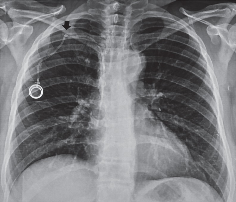
The use of port central venous catheters (CVCs) for chemotherapeutical use has seen exponential growth over the last decades. However, port CVC misplacement may lead to catheter malfunction (such as partial or total catheter blockade), which might be complicated by thrombosis and catheter superinfections, and these in turn may lead to pulmonary embolism and bloodstream infections. The overall occurrence of port CVC misplacement is up to 6%; nonetheless, port CVC misplacement may occur in up to 67% of patients with early CVC dysfunction (occurring within three months of catheter insertion). Thereafter, the prompt evaluation of catheter position among patients with first-trimester CVC dysfunction is extremely important. The aim of the present manuscript is to support medical oncologists, haematologists, and clinicians in timely suspicion and recognition of port CVC misplacement among patients with early CVC dysfunction. Radiological educational iconographic materials that will assist a prompt estimate of port-CVC dislocation are provided.

Keywords: CVCs; central venous catheters; chemotherapy; dysfunction; misplacement; port.


Conflict of interest statement

The authors declare no conflict of interest.

Figures

Fig. 1
Fig. 1
Correct placement. Catheter tip terminates in the superior vena cava, just upstream of the right atrium
Fig. 2
Fig. 2
Correct placement. Catheter tip terminates in the superior vena cava, just upstream of the right atrium
Fig. 3
Fig. 3
Catheter kinked due to compression between the first rib and the clavicle, thus resulting in pinch-off syndrome
Fig. 4
Fig. 4
Long catheter with catheter tip reaching the inferior vena cava
Fig. 5
Fig. 5
Catheter makes a loop in the ipsilateral jugular vein
Fig. 6
Fig. 6
Catheter makes a loop in the ipsilateral jugular vein
Fig. 7
Fig. 7
Catheter is too long and ends in the contralateral subclavian vein
Fig. 8
Fig. 8
Catheter has entered the right subclavian vein and then continues to the right internal jugular vein
Fig. 9
Fig. 9
Tight suture generating a catheter kinking right after the port. As a result, there is a mechanical obstruction that blocks flow in any direction
Fig. 10
Fig. 10
Tight suture generating a catheter kink right after the port. We can also see catheter length with catheter tip misplaced and reaching the inferior vena cava
Fig. 11
Fig. 11
Chest X-ray indicates a long catheter with endocardial loop in the right atrium
Fig. 12
Fig. 12
The chest X-ray shows a catheter fracture in the right atrium. The fragment must be removed immediately to avoid life-threatening complications. We can also see a pinch-off syndrome due to catheter compression between the first rib and the clavicle
Fig. 13
Fig. 13
The chest X-ray (profile view) shows a catheter fracture in the right atrium
Fig. 14
Fig. 14
Short catheter with catheter tip just at the entrance of the right subclavian vein. There is an extremely high risk of drug extravasation with minimal catheter back movement/migration
Fig. 15
Fig. 15
Short catheter: due to its shortness the catheter has migrated outside the superior vena cava
Fig. 16
Fig. 16
Catheter length with a narrow angle loop immediately after the port and a second narrow angle distortion before its entrance in the subclavian vein
Fig. 17
Fig. 17
Catheter length with a narrow angle loop immediately after the port
Fig. 18
Fig. 18
Flow chart with suggestions on how to manage port-catheter misplacement

Similar articles

Cited by

References

    1. Sousa B, Furlanetto J, Hutka M, Gouveia P, Wuerstlein R, Mariz JM, Pinto D, Cardoso F, ESMO Guidelines Committee Central venous accesss in Oncology: ESMO Clinical Practice Guidelines. Ann Oncol. 2015;26(Suppl. 5):152–168. - PubMed
    1. Schiffer CA, Mangou PB, Wade JC, et al. Central Venous Catheter care for the patient with cancer: American Society of Clinical Oncology Clinical Practice Guideline. J Clin Oncol. 2013;17:1357–1370. - PubMed
    1. Granic M, Zdravkovic D, Krstajic S, et al. Totally implantable central venous catheters of the port-a-cath type: complications due to its use in the treatment of cancer patients. J BUON. 2014;19:842–846. - PubMed
    1. Mauri D, Roumbkou S, Michalopoulou S, Tsali L, et al. Port central venous catheters associated bloodstream infection during outpatient-based chemotherapy. Med Oncol. 2010;27:1309–1313. - PubMed
    1. Wiegering V, Schmid S, Andres O, et al. Thrombosis as a complication of central venous access in pediatric patients with malignancies: a 5-year single-center experience. BMC Hematol. 2014;14:18. - PMC - PubMed

LinkOut - more resources