. 2018 Jul 29:2018:9787128.
doi: 10.1155/2018/9787128. eCollection 2018.

Insulin-Like Growth Factor-I as an Effector Element of the Cytokine IL-4 in the Development of a Leishmania major Infection

Affiliations

Insulin-Like Growth Factor-I as an Effector Element of the Cytokine IL-4 in the Development of a Leishmania major Infection

Luiza C Reis et al. Mediators Inflamm. .

Abstract

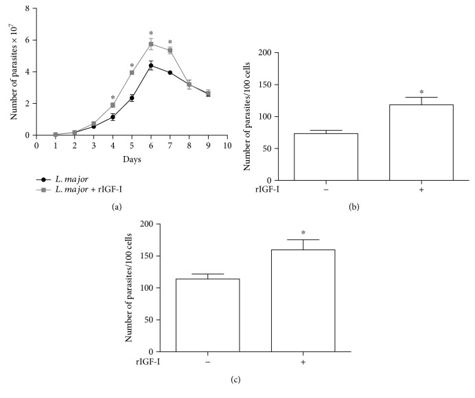
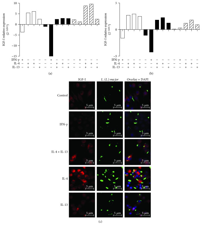
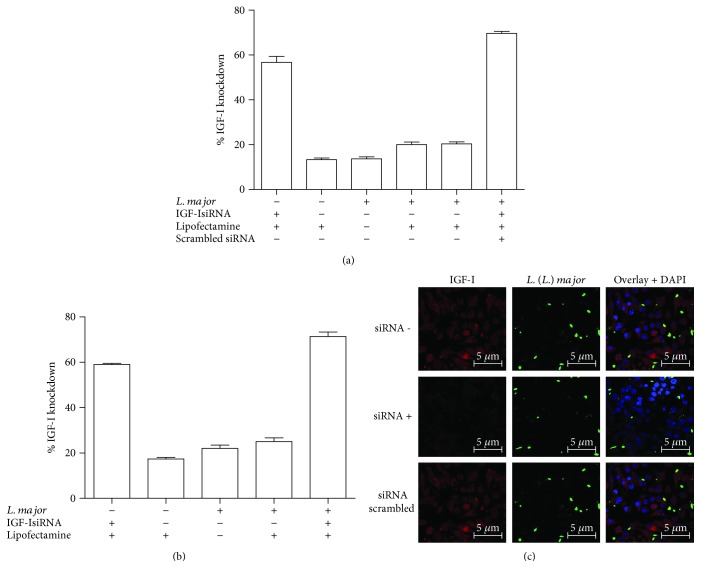
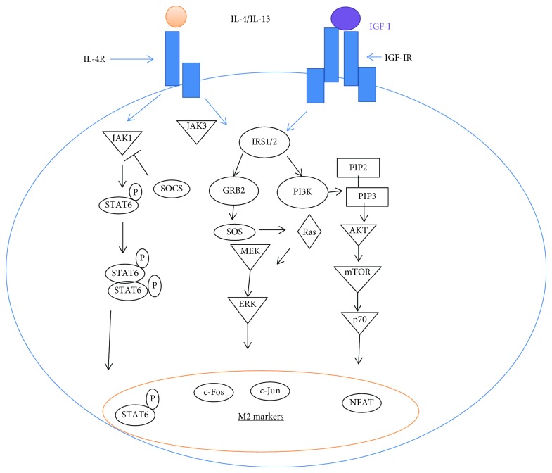
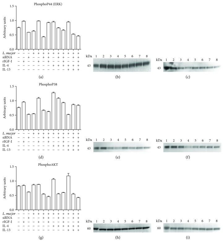
Certain cytokines modulate the expression of insulin-like growth factor- (IGF-) I. Since IL-4 and IGF-I promote growth of the protozoan Leishmania major, we here addressed their interaction in downregulating the expression of Igf-I mRNA using small interfering RNA (siRNA) in Leishmania major-infected macrophages. Parasitism was decreased in the siRNA-treated cells compared with the nontreated cells, reversed by the addition of recombinant IGF-I (rIGF-I). In IL-4-stimulated macrophages, parasitism and the Igf-I mRNA amount were increased, and the effects were nullified upon siRNA transfection. IGF-I downregulation inhibited both parasite and macrophage arginase activation even in IL-4-stimulated cells. Searching for intracellular signaling components shared by IL-4 and IGF-I, upon siRNA transfection, phosphorylated p44, p38, and Akt proteins were decreased, affecting the phosphatidylinositol-3-kinase (PI3K)/Akt pathway. In L. major-infected C57BL6-resistant mice, the preincubation of the parasite with rIGF-I changed the infection profile to be similar to that of susceptible mice. We conclude that IGF-I constitutes an effector element of IL-4 involving the PI3K/Akt pathway during L. major infection.


Figures

Figure 1
Figure 1
The effects of IGF-I on the growth of L. major in culture and within macrophages (mean ± standard deviation). (a) The effect of IGF-I on the growth of Leishmania promastigotes in culture maintained in 199 medium supplemented with 2% heat-inactivated FCS at 26°C is shown. Leishmania promastigotes were maintained with (gray line) or without (black line) 50 ng/mL rIGF-I. (b and c) The effect of IGF-I on the growth of Leishmania within macrophages is shown. RAW 264.7 cells were infected with L. major promastigotes (b) or amastigotes (c) treated with or without 50 ng/mL rIGF-I and incubated for 48 h. P < 0.05 (one-way ANOVA and Student's t test) compared to the control without rIGF-I.
Figure 2
Figure 2
IGF-I expression in response to cytokine treatment. (a and b) Quantification of Igf-I mRNA in L. major promastigote- (black bars) or amastigote-infected (striped bars) or noninfected (white bars) RAW 264.7 cells (a) and BALB/c peritoneal macrophages (b) is shown. Cells were stimulated with IFN-γ (200 U/mL), IL-4 (2 ng/mL), and IL-13 (5 ng/mL) for 48 hours. One representative experiment from three independent assays is shown. (c) Detection of IGF-I expression using confocal microscopy of cells labeled with a 1 : 75 dilution of an anti-IGF-I antibody (using an Alexa Fluor 546-conjugated secondary antibody; red) and a 1 : 200 dilution of an anti-Leishmania antibody (using an Alexa Fluor 488-conjugated secondary antibody; green) in L. major promastigote-infected macrophages is shown. Nuclei were stained with DAPI (blue). Images were captured using a confocal Leica LSM510 microscope with a 63x oil immersion objective. The expressions are relative to the expression in untreated cells (the baseline).
Figure 3
Figure 3
Expression of Igf-I mRNA upon IGF-I silencing with an siRNA. The percentage decrease in the amount of Igf-I mRNA in RAW 264.7 cells (a) or BALB/c peritoneal macrophages (b) infected with L. major promastigotes that were transfected with 150 μM siRNA, scrambled siRNA, or Lipofectamine alone 6 h after infection is shown. One representative experiment from three independent assays is shown. (c) The detection of IGF-I expression in L. major promastigote-infected RAW 264.7 cells transfected with (siRNA+) or without the IGF-I siRNA (siRNA−) or with a scrambled siRNA using confocal microscopy of immunostaining with a 1 : 75 dilution of an anti-IGF-I antibody (using an Alexa Fluor 546-conjugated secondary antibody; red) and a 1 : 200 dilution of an anti-Leishmania antibody (using an Alexa Fluor 488-conjugated secondary antibody; green) is shown. Nuclei were stained with DAPI (blue). Images were captured using a confocal Leica LSM510 microscope with a 63x oil immersion objective.
Figure 4
Figure 4
Parasitism in response to cytokine treatments and siRNA transfection. Parasitism (median number of parasites per 100 cells) in L. major-infected RAW 264.7 cells (a, b, e, and f) or BALB/c peritoneal macrophages (c and d) following transfection with IGF-I siRNA (a, b, c, and d), Lipofectamine alone (e), or scrambled siRNA (f) along with cytokine stimulation is shown. Promastigote- (a, c, e, and f) or amastigote-infected (b and d) cells transfected with or without siRNA or Lipofectamine were stimulated with IFN-γ (200 U/mL), IL-4 (2 ng/mL), IL-13 (5 ng/mL), and recombinant IGF-I (rIGF-I, 50 ng/mL) for 48 hours. One representative experiment from three independent assays is shown. P < 0.05 (ANOVA and Tukey's tests).
Figure 5
Figure 5
The effects of IGF-I siRNA transfection on the L-arginine metabolic pathway. The quantification of (a) Arg1 mRNA, (b) Larg mRNA, (c) Cat-2B mRNA, (d) arginase activity, (e) Nos2 mRNA, and (f) NO production in L. major-infected RAW 264.7 cells following transfection with IGF-I siRNA and cytokine stimulation is shown. Promastigote-infected cells transfected with or without IGF-I siRNA were stimulated with IFN-γ (200 U/mL), IL-4 (2 ng/mL), and IL-13 (5 ng/mL) for 48 hours. One representative experiment from three independent assays is shown. P < 0.05 (ANOVA and Tukey's tests) compared to the respective control without siRNA.
Figure 6
Figure 6
Scheme of the common components of the IGF-I and IL-4 signaling pathways.
Figure 7
Figure 7
The effects of siRNA and IL-4 on components of the IGF-I signaling pathways: levels of phosphorylated p44 (ERK), p38 (MAPK), and AKT proteins. Promastigote-infected or noninfected cells transfected with or without IGF-I siRNA were stimulated for 30 minutes with IL-4 (2 ng/mL) and IL-13 (5 ng/mL). Cells were lysed, proteins were separated by 10% SDS-PAGE, and subsequently, Western blotting was performed using anti-phospho-p44 (a, b, and c), anti-phospho-p38 (d, e, and f), and anti-phospho-AKT (g, h, and i) antibodies. Protein bands corresponding to protein expression levels were subject to a densitometric analysis, and the data are expressed in arbitrary units (a, d, and g). A representative blot is shown. (b, e, h) The lanes represented the following: 1: control; 2: RAW; 3: RAW + rIGF; 4: RAW + siRNA; 5: RAW + Lm; 6: RAW + Lm + rIGF; 7: RAW + Lm + IL-4; and 8: RAW + Lm + IL-4 + IL-13. (c, f, i): 1: control; 2: RAW; 3: RAW + Lm + siRNA; 4: RAW + Lm + siRNA + rIGF; 5: RAW + Lm + siRNA + IL-4; 6: RAW + Lm + siRNA + IL-4 + rIGF; 7: RAW + Lm + siRNA + IL-4 + IL-13; and 8: RAW + Lm + siRNA + IL-4 + IL-13 + rIGF. See Materials and Methods for additional details.
Figure 8
Figure 8
IGF-I expression and the effect of IGF-I on lesion development in L. major-susceptible and -resistant mouse strains. (a) The ratio of the Igf-I mRNA amount to β-actin mRNA amount in L. major-infected BALB/c (white bars) and C57BL/6 peritoneal macrophages (black bars) is shown. (b) Confocal microscopy was used to detect IGF-I expression via immunostaining with a 1 : 75 dilution of an anti-IGF-I antibody (using an Alexa Fluor 546-conjugated secondary antibody; red) and a 1 : 200 dilution of an anti-Leishmania antibody (using an Alexa Fluor 488-conjugated secondary antibody; green). DAPI (blue) was used to stain the nuclei. Images were captured using a confocal Leica LSM510 microscope with a 63x oil immersion objective. (c and d) Stationary-phase promastigotes (106) that were preincubated with or without recombinant IGF-I (50 ng/mL) for 5 min were injected into the footpads of BALB/c and C57BL/6 mice, and lesion development was measured for six weeks. One representative experiment from three independent assays is shown. P < 0.05 (ANOVA and Tukey's tests) compared to the respective controls. #P < 0.05 (ANOVA and Tukey's tests) between BALB/c and C57BL/6.

References

    1. Frystyk J. Free insulin-like growth factors -- measurements and relationships to growth hormone secretion and glucose homeostasis. Growth Hormone & IGF Research. 2004;14(5):337–375. doi: 10.1016/j.ghir.2004.06.001. - DOI - PubMed
    1. Hehlgans T., Pfeffer K. The intriguing biology of the tumour necrosis factor/tumour necrosis factor receptor superfamily: players, rules and the games. Immunology. 2005;115(1):1–20. doi: 10.1111/j.1365-2567.2005.02143.x. - DOI - PMC - PubMed
    1. Valverde A. M., Benito M., Lorenzo M. The brown adipose cell: a model for understanding the molecular mechanisms of insulin resistance. Acta Physiologica Scandinavica. 2005;183(1):59–73. doi: 10.1111/j.1365-201X.2004.01384.x. - DOI - PubMed
    1. Arkins S., Rebeiz N., Brunke-Reese D. L., Biragyn A., Kelley K. W. Interferon-gamma inhibits macrophage insulin-like growth factor-I synthesis at the transcriptional level. Molecular Endocrinology. 1995;9(3):350–360. doi: 10.1210/mend.9.3.7776981. - DOI - PubMed
    1. O'Connor J. C., McCusker R. H., Strle K., Johnson R. W., Dantzer R., Kelley K. W. Regulation of IGF-I function by proinflammatory cytokines: at the interface of immunology and endocrinology. Cellular Immunology. 2008;252(1-2):91–110. doi: 10.1016/j.cellimm.2007.09.010. - DOI - PMC - PubMed

MeSH terms

Substances

LinkOut - more resources