. 2018 Aug 13:9:1865.
doi: 10.3389/fimmu.2018.01865. eCollection 2018.

Interleukin-6 and Type-I Collagen Production by Systemic Sclerosis Fibroblasts Are Differentially Regulated by Interleukin-17A in the Presence of Transforming Growth Factor-Beta 1

Affiliations

Interleukin-6 and Type-I Collagen Production by Systemic Sclerosis Fibroblasts Are Differentially Regulated by Interleukin-17A in the Presence of Transforming Growth Factor-Beta 1

Aleksandra Maria Dufour et al. Front Immunol. .

Abstract

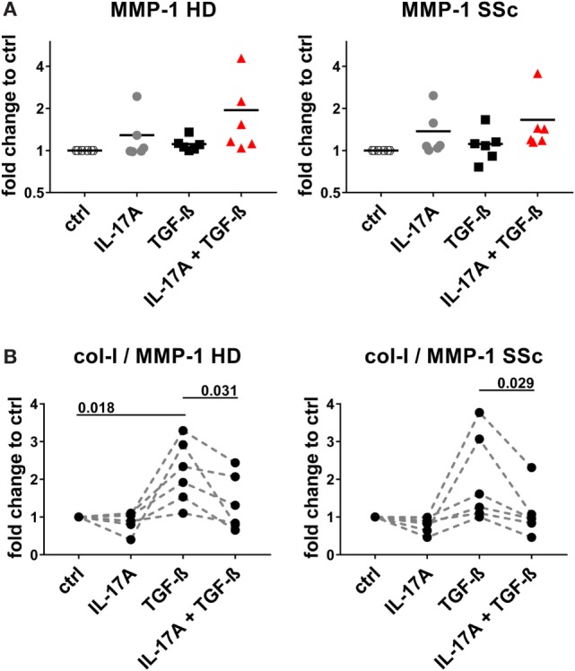
Functional cytokine networks have been poorly characterized in systemic sclerosis (SSc). While interleukin-17A (IL-17A) is increased in SSc skin and other organs, its role is still debated, particularly considering fibrogenesis. We uncover here a dual function of IL-17A in the presence of transforming growth factor-β 1 (TGF-β), the master pro-fibrotic cytokine. In the one hand, we report an unexpected synergic activity resulting in enhanced production of IL-6 by dermal fibroblasts; in the other hand, a substantial inhibition of type I collagen (col-I) production. IL-17A or TGF-β enhanced the production of IL-6 by 8- to 16-folds when compared to control in healthy donors (HD) and SSc cultures. However, the joint presence of IL-17A and TGF-β resulted in robustly exuberant responses with levels of IL-6 up to 100-folds higher than those observed in untreated cells. Inhibition of NFκB signaling pathway preferentially inhibited the production of IL-6 driven by IL-17A in HD fibroblasts, while inhibition of PI3K preferentially inhibited the production of IL-6 driven by TGF-β. Interestingly, when p38 MAPK was inhibited, substantial reduction of IL-6 production was observed for both IL-17A and TGF-β. Consistently with the inhibition experiments, the combined stimulation of fibroblasts by IL-17A and TGF-β resulted in 1.8-fold increase in p38 MAPK phosphorylation (P = 0.025), when compared to levels of phosphorylated p38 MAPK induced by IL-17A alone. Furthermore, the enhanced phosphorylation of p38 MAPK in the joint presence of IL-17A and TGF-β was unique among the signaling molecules we examined. As expected, TGF-β induced SMAD2 phosphorylation and col-I production. However, in fibroblasts cultured in the joint presence of TGF-β and IL-17A, SMAD2 phosphorylation was decreased by 0.6-folds (P = 0.022) when compared to that induced by TGF-β alone. Remarkably, in this condition, the production of col-I and fibronectin was significantly decreased in both HD and SSc. Thus, IL-17A and TGF-β reciprocally influence each other effector functions in fibroblasts. Intracellular molecular switches may favor synergic or antagonistic activities, which are revealed by specific readouts. The implications of these data in the context of SSc are far reaching, particularly in terms of therapeutic approaches since IL-6, IL-17A, and TGF-β are all putative targets of treatment.

Keywords: fibrosis; interleukin-17A; interleukin-6; monocyte chemotactic protein-1; systemic sclerosis; transforming growth factor-beta; type-I collagen.


Figures

Figure 1
Figure 1
IL-6 and monocyte chemotactic protein-1 (MCP-1) are synergistically and specifically induced in human fibroblasts by the combined action of IL-17A and TGF-β. Primary human dermal fibroblasts from healthy donors (HD) and systemic sclerosis (SSc) patients were cultured in the presence of IL-17A (25 ng/ml), TGF-β (2.5 ng/ml), or their combination for 48 h, in 96-well plates, in triplicates. IL-6 (A), MCP-1 (B), IL-8 (C) were assessed by ELISA in culture supernatants. Results are expressed as fold change compared to spontaneous production in control (ctrl) cultures. Basal levels were: 22.7 (±7.3) and 40.7 (±16.7) pg/ml for IL-6; 328.1 (±33.3) and 377.2 (±85.4) pg/ml for MCP-1 and 211.8 (±56.6) and 207.7 (±56.9) pg/ml for IL-8, in HD and SSc, respectively. Significance was assessed by paired t test.
Figure 2
Figure 2
TGF-β inhibition abrogates the synergistic response with IL-17A. HD fibroblasts were treated with (A) 10 µg/ml TGF-β 1 neutralizing antibody or 10 µg/ml of an irrelevant ctrl Ab, (B) SD208 (TGFβR1 inhibitor) or vehicle for 1 h prior to the addition of IL-17A (25 ng/ml) or TGF-β (2.5 ng/ml) and cultured for 48 h. IL-6 levels in SN were assessed by ELISA. Results are shown as fold change to untreated control cultures (basal level of IL-6 was 3.1 ± 1.1 pg/ml). (B) Square: TGF-β (10 ng/ml); empty circle: (SD2018, 1 µM); full circle: vehicle. Significant differences were assessed by paired t-test. Bars in (A) and symbols in (B) represent the mean + SEM of three experiments.
Figure 3
Figure 3
Shared and private signaling pathways are preferentially used by IL-17A and TGF-β to induce IL-6. Healthy donors fibroblasts were treated with optimal doses of inhibitors (20 µM U0126, 20 µM SB203580, 10 µM SP600125, 10 µM Ly294002, or 0.37 µM TPCA-1) or vehicle for 1 h prior to the addition of IL-17A (25 ng/ml) or TGF-β (2.5 ng/ml) and cultured for an additional 48 h, in triplicates. (A) IL-6 levels in SN were assessed by ELISA. Results are shown as the percentage of IL-6 production induced by IL-17A or TGF-β in the absence of inhibitors (levels of IL-6 were: 22.8 ± 3.3 pg/ml for IL-17A and 8.8 ± 3.9 pg/ml for TGF-β). Bars represent the mean + SEM of three experiments. Significant differences versus control were assessed by paired t-test: *P < 0.05, ****P < 0.001. (B). Fibroblast viability was assessed by EZ4U and found >90% for all culture conditions.
Figure 4
Figure 4
NFκB and PI3K signaling pathways are preferentially used by IL-17A and TGF-β, respectively and together cooperate in inducing IL-6. Healthy donors fibroblasts were treated with the indicated concentrations of (A) TPCA-1; (B) Ly294002; or (C) suboptimal doses of TPCA-1 (0.03 µM) and/or Ly294002 (Ly, 2 µM) for 1 h prior to addition of IL-17A (25 ng/ml) and/or TGF-β (2.5 ng/ml). After 48 h, culture SNs were collected and IL-6 levels were assessed by ELISA. (A,B) Results are shown as fold change to untreated cells, mean + SEM is indicated (N = 4). Please note the log2 scale. (C) Results are shown as the percentage of IL-6 production induced by IL-17A and/or TGF-β in the absence of inhibitors (levels of IL-6 were: 86.2 ± 11.8 pg/ml for IL-17A, 49.6 ± 10.2 pg/ml for TGF-β, and 257.5 ± 82.5 pg/ml for IL-17A + TGF-β). Bars represent the mean + SEM. Significant differences versus control were assessed by paired t-test: *P < 0.05, **P < 0.01, ***P < 0.001, ****P < 0.001.
Figure 5
Figure 5
p38 MAPK signaling pathway is common to IL-17A- and TGF-β-induced IL-6 production. Healthy donors fibroblasts were treated with the indicated concentrations of SB203580 (A) or 20 µM SB203580 (B) for 1 h prior to the addition of IL-17A (25 ng/ml) and/or TGF-β (2.5 ng/ml) in triplicates. After 48 h, culture SNs were collected and IL-6 levels were assessed by ELISA. (A) Results are shown as fold change to untreated cells, mean + SEM is indicated, N = 3. Please note the log2 scale. (B) Results are shown as the percentage of IL-6 production induced by IL-17A and/or TGF- β in the absence of inhibitor (levels of IL-6 were: 14.7 ± 7.4 pg/ml for IL-17A, 11.4 ± 6.1 pg/ml for TGF-β, and 48.9 ± 13.7 pg/ml for IL-17A + TGF-β). Bars represent the mean + SEM of three experiments. Significant differences versus control were assessed by paired t-test.
Figure 6
Figure 6
Phosphorylation of MAPK p38 is enhanced by the combined action of IL-17A and TGF-β. (A) Western blot (WB) of healthy donors fibroblasts treated with 1 µM TPCA-1 and/or 10 µM Ly294002 for 1 h prior to addition of IL-17A (25 ng/ml) and/or TGF-β (2.5 ng/ml) and cultured for an additional 10 min. Results are representative of three experiments with inhibitors and two additional experiments with cytokines only. (B) Quantification of Western blot (WB) analysis was performed with ImageJ software (http://rsbweb.nih.gov/ij) and values were normalized to β-actin, N = 5. Results are shown as fold change to IL-17A-treated cells (for p-p38, p-IκBα, and p-NF-κB p65) or to untreated cells (for p-Akt), N = 5. Significance assessed by paired t test.
Figure 7
Figure 7
IL-17A inhibits SMAD2 phosphorylation and production of type I collagen and fibronectin induced by TGF-β. Healthy donors (HD) fibroblasts were treated with IL-17A (25 ng/ml) and/or TGF-β (2.5 ng/ml) for 10 min. (A). Western blot (WB) representative of five distinct experiments. (B) Quantification of WB analysis. Results are shown as fold change to TGF-β-treated cells, N = 5. (C,D) Primary human fibroblasts from HD (left panel) and systemic sclerosis (SSc) patients (right panel) in triplicates were treated with IL-17A (25 ng/ml), TGF-β (2.5 ng/ml), or a combination of these cytokines. After 48 h, culture supernatants were collected and type I collagen (C) and fibronectin (D) levels were assessed by ELISA (N = 5). Results are shown as fold change to untreated cells. Basal levels for col-I were 364.8 (±91.0) and 203.8 (±66.5) ng/ml and for fibronectin 972.7 (±273.9) and 1036,4 (±307.6) ng/ml, in HD and SSc, respectively. Significance was assessed by paired t-test.
Figure 8
Figure 8
IL-17A decreases the col-I to MMP-1 ratio enhanced by TGF-β. Primary human dermal fibroblasts from healthy donors (HD) (left panel) and systemic sclerosis (SSc) patients (right panel) were cultured in the presence of IL-17A (25 ng/ml), TGF-β (2.5 ng/ml), or their combination for 48 h, in 96-well plates, in triplicates. MMP-1 levels (A) were assessed by ELISA in culture supernatants. Results are expressed as fold change compared to spontaneous production in control (ctrl) cultures. Basal levels for MMP-1 were 15.71 (±1.3) and 20.1 (±2.3) ng/ml, in HD and SSc, respectively. (B) The ratio of col-I levels from Figure 7C to MMP-1 was calculated. Significance was assessed by paired t-test.
Figure 9
Figure 9
Proposed model linking IL-17A, TGF-β, and IL-6 in the context of extracellular matrix deposition and Th17 cell differentiation. Blue arrows: stimulatory signal; red arrows: inhibitory signal. The relevant references are reported in the discussion. For tissue inhibitor of metalloproteinases 1 (TIMP-1), we refer to Fineschi et al. (45).

References

    1. Gabrielli A, Avvedimento EV, Krieg T. Scleroderma. N Engl J Med (2009) 360(19):1989–2003.10.1056/NEJMra0806188 - DOI - PubMed
    1. Varga JA, Trojanowska M. Fibrosis in systemic sclerosis. Rheum Dis Clin North Am (2008) 34(1):115–43.10.1016/j.rdc.2007.11.002 - DOI - PMC - PubMed
    1. Allanore Y, Simms R, Distler O, Trojanowska M, Pope J, Denton CP, et al. Systemic sclerosis. Nat Rev Dis Primers (2015) 1:15002.10.1038/nrdp.2015.2 - DOI - PubMed
    1. Chizzolini C, Brembilla NC, Montanari E, Truchetet ME. Fibrosis and immune dysregulation in systemic sclerosis. Autoimmun Rev (2011) 10(5):276–81.10.1016/j.autrev.2010.09.016 - DOI - PubMed
    1. Wynn TA, Ramalingam TR. Mechanisms of fibrosis: therapeutic translation for fibrotic disease. Nat Med (2012) 18(7):1028–40.10.1038/nm.2807 - DOI - PMC - PubMed

Publication types