

Case Reports
doi: 10.3109/01658107.2011.559683.
eCollection 2011.
Idiopathic Hypertrophic Cranial Pachymeningitis Associated With Intermediate Uveitis
Affiliations
- PMID: 30151029
- PMCID: PMC6104769
- DOI: 10.3109/01658107.2011.559683
Item in Clipboard
Case Reports
Idiopathic Hypertrophic Cranial Pachymeningitis Associated With Intermediate Uveitis
Neuroophthalmology.
.
Abstract
The authors report a case with idiopathic hypertrophic cranial pachymeningitis associated with intermediate uveitis. The patient complained of decreased vision in both eyes, especially the right. Ophthalmic examination revealed right optic disc pallor, bilateral vitritis, and cystoid macular edema. Magnetic resonance imaging revealed marked enhancement of a dural lesion. The macular edema responded well to medical treatment. Intermediate uveitis has not yet been reported in the context of idiopathic hypertrophic cranial pachymeningitis.
Keywords: cystoid macular oedema; idiopathic hypertrophic cranial pachymeningitis; intermediate uveitis.
Figures

Fluorescein angiography images show cystoid macular oedema (arrows) and peripheral vascular leakage (arrowheads) (a, b, c). One month later images show resolution of macular oedema and a decrease in the peripheral vascular leakage (d, e, f).
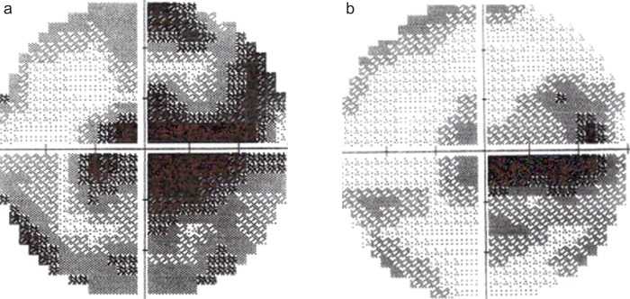
Humphrey visual field test shows central scotoma, enlargement of the blind spot, and peripheral scotoma in the right eye (a). The test performed 2 weeks later showed partial regression of scotomas (b).

Coronal post-gadolinium image performed in April 2008 shows the lesion extending from right cavernous sinus to the prechiasmatic part of right optic nerve (a, arrow). Axial post-gadolinium image of the brain 9 years after diagnosis shows residual thickening of the tentorium cerebelli (on prednisone and azathioprine therapy) (b, arrow).
Similar articles
-
A Rare Ocular Manifestation of Idiopathic Hypertrophic Cranial Pachymeningitis.Cureus. 2021 Dec 23;13(12):e20633. doi: 10.7759/cureus.20633. eCollection 2021 Dec. Cureus. 2021. PMID: 35106199 Free PMC article.
-
Etanercept monotherapy induces complete resolution of cystoid macular edema in a patient with intermediate uveitis.Retin Cases Brief Rep. 2007 Fall;1(4):261-3. doi: 10.1097/01.iae.0000242843.02952.46. Retin Cases Brief Rep. 2007. PMID: 25390997
-
Extensive anterior cranial fossa idiopathic hypertrophic pachymeningitis: a case report and review of the literature.J Med Assoc Thai. 2005 Dec;88(12):1934-40. J Med Assoc Thai. 2005. PMID: 16518996 Review.
-
Idiopathic hypertrophic cranial pachymeningitis associated with total occlusion of the dural sinuses--case report.Neurol Med Chir (Tokyo). 2004 Dec;44(12):650-4. doi: 10.2176/nmc.44.650. Neurol Med Chir (Tokyo). 2004. PMID: 15684597
-
Idiopathic hypertrophic cranial pachymeningitis. Report of three cases.J Neurosurg. 1993 Aug;79(2):270-6. doi: 10.3171/jns.1993.79.2.0270. J Neurosurg. 1993. PMID: 8331412 Review.
Cited by
-
Idiopathic hypertrophic pachymeningitis in a patient with a history of diffuse large B cell lymphoma.BMJ Case Rep. 2023 Jun 14;16(6):e254847. doi: 10.1136/bcr-2023-254847. BMJ Case Rep. 2023. PMID: 37316284 Free PMC article.
-
A Rare Ocular Manifestation of Idiopathic Hypertrophic Cranial Pachymeningitis.Cureus. 2021 Dec 23;13(12):e20633. doi: 10.7759/cureus.20633. eCollection 2021 Dec. Cureus. 2021. PMID: 35106199 Free PMC article.
-
Recurrent idiopathic hypertrophic pachymeningitis with chronic headache and cranial nerve involvement.Proc (Bayl Univ Med Cent). 2024 Feb 8;37(2):322-325. doi: 10.1080/08998280.2023.2298654. eCollection 2024. Proc (Bayl Univ Med Cent). 2024. PMID: 38343488 Free PMC article.
References
-
- Kupersmith M.J, Martin V, Heler G, Shah A, Mitnick HJ. Idiopathic hypertrophic pachymeningitis. Neurology 2004;62:686–694. - PubMed
-
- Aıldere AM, Tutar NU, Yücel E, Coşkun M, Benli S, Aydin P. Case report. Pachymeningitis and optic neuritis in rheumatoid arthritis. MRI findings. Br J Radiol 1999;72:404–407. - PubMed
-
- Specks U, Moder K, McDonald T. Meningeal involvement in Wegener granulomatosis. Mayo Clin Proc 2000;75:856–859. - PubMed
-
- Yamashita K, Suzuki Y, Yoshizumi H, Takahashi JB, Nogawa T. Tuberculous hypertrophic pachymeningitis involving the posterior fossa and high cervical region-case report. Neurol Med Chir (Tokyo) 1994;34:100–103. - PubMed
-
- Nussenblatt RB, Whitcup SM. Uveitis: Fundamentals and Clinical Practice. 3th ed Philadelphia: Elsevier, 2004;291–300.
Publication types
LinkOut - more resources
Full Text Sources