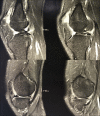
Anterior and posterior cruciate ligament agenesis
- PMID: 30151108
- PMCID: PMC6101568
- DOI: 10.1093/jscr/rjy216
Anterior and posterior cruciate ligament agenesis
Abstract
Congenital absence of the cruciate ligament is a rare condition with a prevalence of 0.017 per 1000 live births. This study reports a case of congenital absence of the anterior and posterior cruciate ligaments of the left knee associated to a type 1A fibular hemimelia, and a contribution to the existing hypotheses on knee ligaments development. According to medical literature the anomaly begins to develop around the seventh-eighth week of pregnancy. Patients with a cruciate ligament agenesis will often need a knee replacement at one point in their lives.
Figures


References
-
- Johansson E, Aparisi T. Congenital absence of the cruciate ligaments: a case report and review of the literature. Clin Orthop Relat Res 1982;162:108–11. - PubMed
-
- Manner HM, Radler C, Ganger R, Grill F. Dysplasia of the cruciate ligaments: radiographic assessment and classification. J Bone Joint Surg Am 2006;88:130–7. - PubMed
-
- Berruto M, Gala L, Usellini E, Duci D, Marelli B. Congenital absence of the cruciate ligaments. Knee Surg Sports Traumatol Arthrosc 2012;20:1622–5. - PubMed
-
- Silva A, Sampaio R. Anterior lateral meniscofemoral ligament with congenital absence of the ACL. Knee Surg Sports Traumatol Arthrosc 2012;20:1622–5. - PubMed
-
- Malchér MG, Bruno AAM, Grisone B, Bernardelli G, Pietrogrande L. Isolated congenital absence of posterior cruciate ligament. A case report. Chir Organi Mov 2008;92:105–7. - PubMed
Publication types
LinkOut - more resources
Full Text Sources
Other Literature Sources

