X-ray computed tomography and its potential in ecological research: A review of studies and optimization of specimen preparation
- PMID: 30151184
- PMCID: PMC6106166
- DOI: 10.1002/ece3.4149
X-ray computed tomography and its potential in ecological research: A review of studies and optimization of specimen preparation
Abstract
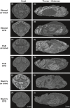
Imaging techniques are a cornerstone of contemporary biology. Over the last decades, advances in microscale imaging techniques have allowed fascinating new insights into cell and tissue morphology and internal anatomy of organisms across kingdoms. However, most studies so far provided snapshots of given reference taxa, describing organs and tissues under "idealized" conditions. Surprisingly, there is an almost complete lack of studies investigating how an organism's internal morphology changes in response to environmental drivers. Consequently, ecology as a scientific discipline has so far almost neglected the possibilities arising from modern microscale imaging techniques. Here, we provide an overview of recent developments of X-ray computed tomography as an affordable, simple method of high spatial resolution, allowing insights into three-dimensional anatomy both in vivo and ex vivo. We review ecological studies using this technique to investigate the three-dimensional internal structure of organisms. In addition, we provide practical comparisons between different preparation techniques for maximum contrast and tissue differentiation. In particular, we consider the novel modality of phase contrast by self-interference of the X-ray wave behind an object (i.e., phase contrast by free space propagation). Using the cricket Acheta domesticus (L.) as model organism, we found that the combination of FAE fixative and iodine staining provided the best results across different tissues. The drying technique also affected contrast and prevented artifacts in specific cases. Overall, we found that for the interests of ecological studies, X-ray computed tomography is useful when the tissue or structure of interest has sufficient contrast that allows for an automatic or semiautomatic segmentation. In particular, we show that reconstruction schemes which exploit phase contrast can yield enhanced image quality. Combined with suitable specimen preparation and automated analysis, X-ray CT can therefore become a promising quantitative 3D imaging technique to study organisms' responses to environmental drivers, in both ecology and evolution.
Keywords: anatomy; animal imaging; computed tomography; internal morphology; μ‐CT.
Figures


References
-
- Abel, R. L. , Laurini, C. R. , & Richter, M. (2012). A palaeobiologist's guide to ‘virtual’ micro‐CT preparation. Palaeontologia Electronica, 15, 1–16.
-
- Amossé, J. , Turberg, P. , Kohler‐Milleret, R. , Gobat, J. M. , & Le Bayon, R. C. (2015). Effects of endogeic earthworms on the soil organic matter dynamics and the soil structure in urban and alluvial soil materials. Geoderma, 243–244, 50–57. 10.1016/j.geoderma.2014.12.007 - DOI
-
- Auclerc, A. , Capowiez, Y. , Guérold, F. , & Nahmani, J. (2013). Application of X‐ray tomography to evaluate liming impact on earthworm burrowing activity in an acidic forest soil under laboratory conditions. Geoderma, 202–203, 45–50. 10.1016/j.geoderma.2013.03.011 - DOI
Publication types
Associated data
LinkOut - more resources
Full Text Sources
Other Literature Sources
Research Materials

