. 2018 Jun;2(6):429-442.
doi: 10.1038/s41551-018-0243-9. Epub 2018 Jun 4.

Mechanically induced development and maturation of human intestinal organoids in vivo

Affiliations

Mechanically induced development and maturation of human intestinal organoids in vivo

Holly M Poling et al. Nat Biomed Eng. 2018 Jun.

Abstract

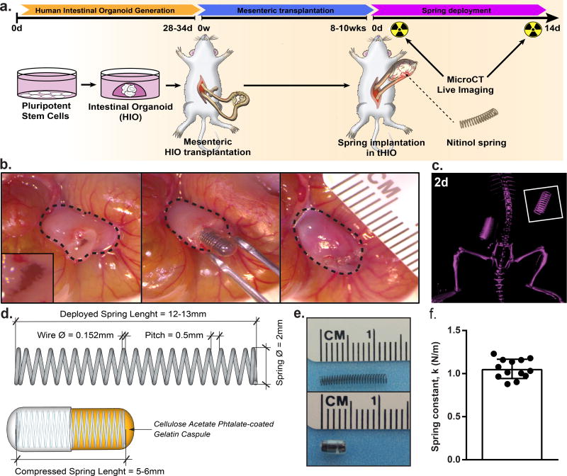
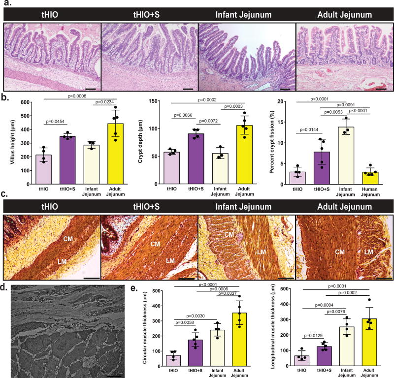
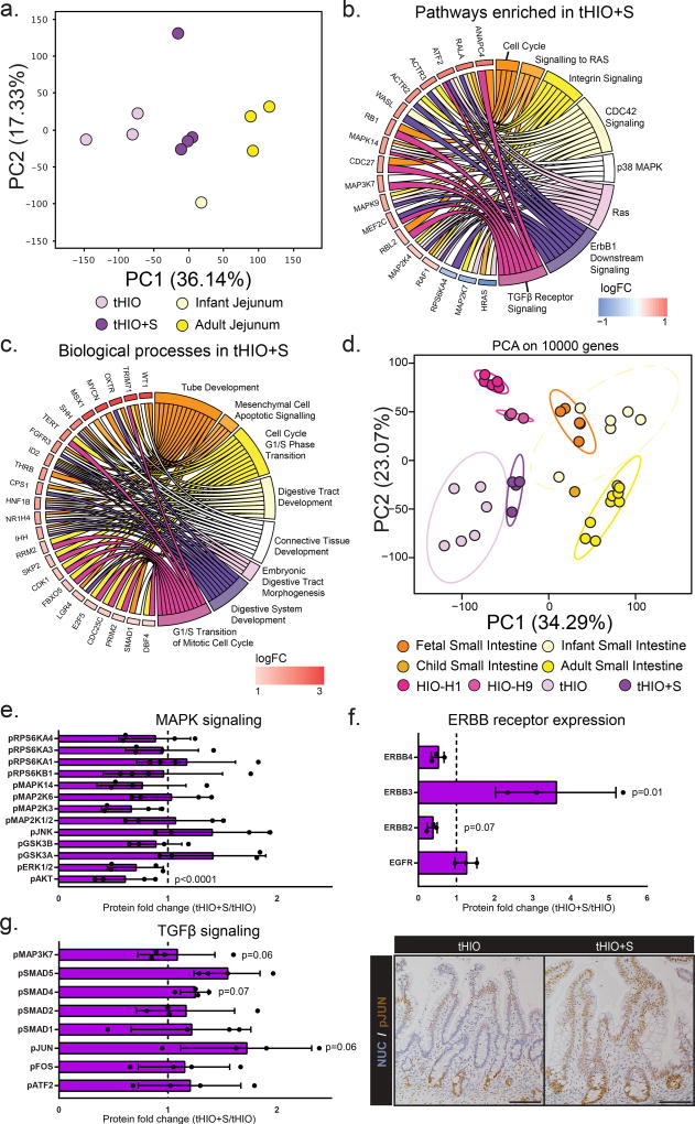
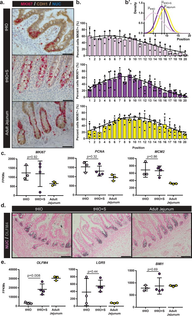
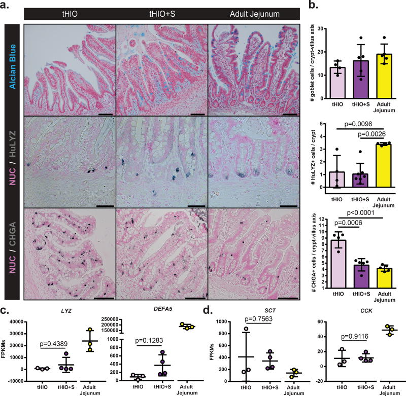
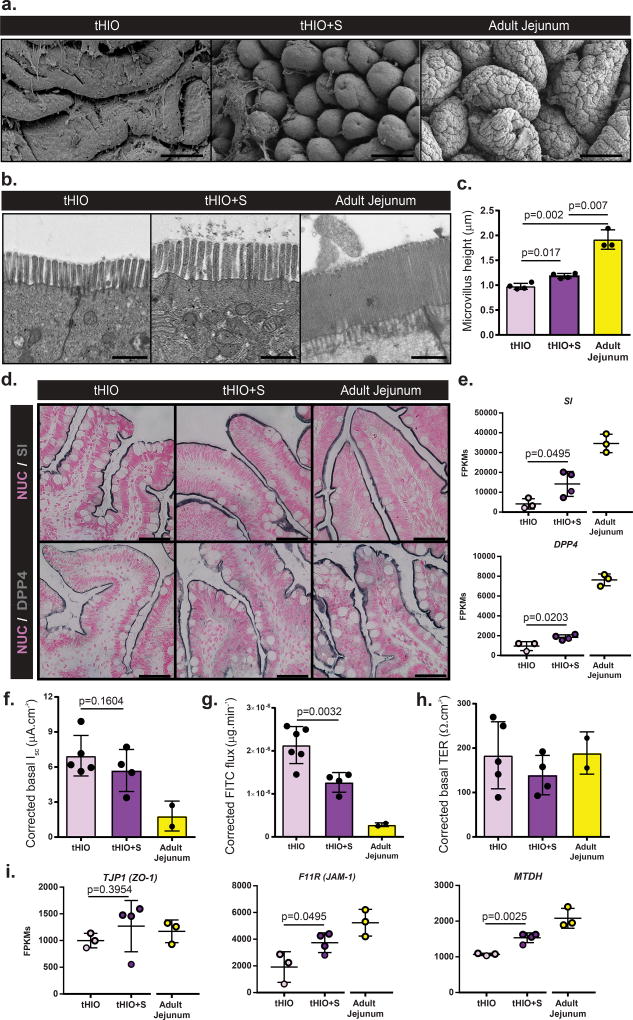
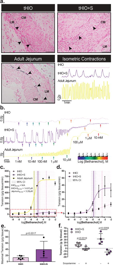
The natural ability of stem cells to self-organize into functional tissue has been harnessed for the production of functional human intestinal organoids. Although dynamic mechanical forces play a central role in intestinal development and morphogenesis, conventional methods for the generation of intestinal organoids have relied solely on biological factors. Here, we show that the incorporation of uniaxial strain, by using compressed nitinol springs, in human intestinal organoids transplanted into the mesentery of mice induces growth and maturation of the organoids. Assessment of morphometric parameters, transcriptome profiling, and functional assays of the strain-exposed tissue revealed higher similarities to native human intestine, with regards to tissue size and complexity, and muscle tone. Our findings suggest that the incorporation of physiologically relevant mechanical cues during the development of human intestinal tissue enhances its maturation and enterogenesis.

Keywords: Human Pluripotent Stem Cells; Small Intestine; Tissue Engineering; Transplantation; Uniaxial Strain.


Figures

Figure 1
Figure 1. Transplantation of springs into tHIOs
(a) 28–34 day old HIOs were transplanted into the mesentery of NSG mice and allowed to grow for 8–10 weeks. Then, a second procedure was performed wherein a compressed NiTi spring was implanted inside the tHIO. Harvest occurred 14 days post spring implantation. (b) Procedural images of the spring insertion into a tHIO. Dashed line indicates perimeter of tHIO. (c) MicroCT of a linearly deployed spring in vivo 2 days after implantation. (d) Schematic of springs used in experiments. Springs utilized for transplantation had a relaxed length of 12–13 mm, compressed length of 5–6 mm and an outside diameter of 2 mm. Compression of springs was maintained through use of a gelatin capsule subsequently coated with a polymer to delay deployment. (e) Photographs of springs used in relaxed (top) and compressed/encapsulated (bottom) forms. (f) The spring constant of those used was 1.05 ± 0.11 N/m (n=13 independent samples). Data are represented as mean ± SD.
Figure 2
Figure 2. tHIO+S samples exhibit increased morphological characteristics
(a) Representative H&E sections of tHIO, tHIO+S, infant jejunum and adult jejunum. Scale bar = 50 µm. This experiment was repeated three times independently and findings were similar. (b) Morphometric quantification of tissue sections was plotted. Villus height, crypt depth and crypt fission were increased in tHIO+S compared to tHIO and better approximate human tissue in the cases of villus height and crypt depth. Sample sizes are the following: tHIO n=4, tHIO+S n=4, infant jejunum n=3, and adult jejunum n=5. All samples are biologically independent. (c) Representative pentachrome sections of tHIO, tHIO+S, infant jejunum and adult jejunum. Scale bar = 100 µm. This experiment was repeated three times independently and findings were similar. (d) Transmission electron micrograph displaying perpendicular orientation of muscle fibers in tHIO+S, similar orientation was observed in tHIO. This experiment was repeated three times independently and findings were similar. (e) Quantification of muscle in pentachrome sections was plotted. Layers of circular and longitudinal muscle (CM; LM respectively) were thicker in tHIO+S compared to tHIO and trend toward that of human jejunum. Sample sizes are the following: tHIO n=4, tHIO+S n=5, infant jejunum n=3, and adult jejunum n=5. All samples are biologically independent. For (b,e) data are represented as the mean ± SD. An ANOVA followed by Tukey’s post hoc tests were performed and the statistical significance cutoff was p < 0.05.
Figure 3
Figure 3. Transcriptionally tHIO+S are matured beyond tHIO
(a) Scaled Centered Principal Component Analysis of the tHIOs, tHIO+S, human infant and adult jejunum was performed. tHIO+S clustered closer to human jejunum tissues. Sample sizes are the following: tHIO n=3, tHIO+S n=4, infant jejunum n=1, and adult jejunum n=3. All samples are biologically independent. (b) Functional enrichment of the pathways upregulated in tHIO+S compared to tHIOs. Sample sizes are the following: tHIO n=3, tHIO+S n=4. All samples are biologically independent. (c) Differential expression gene analysis between tHIO+S and human tissues demonstrated upregulated biological processes in the GO categories concerning intestinal development. Sample sizes are the following: tHIO+S n=4 and adult jejunum n=3. All samples are biologically independent. (d) Scaled Centered Principal Component Analysis of samples retrieved from our study and several publicly available databases. Sample sizes are the following: fetal small intestine n=5, infant small intestine n=6, child small intestine n=1, adult small intestine n=9, HIO-H1 n=5, HIO-H9 n=3, tHIO n=6, and tHIO+S n=4. All samples are biologically independent. (e) Fold-change in MAPK signaling protein array. n=4 biologically independent samples for all groups. (f) Fold-change in ERBB receptor protein expression. n=3 biologically independent samples for all groups. (g) (Left panel) Fold-change in TGFβ signaling protein array. n=4 biologically independent samples for all groups. (Right panel) Immunostaining of phosphorylated JUN in tHIO and tHIO+S. Data are representative of n=4 for all groups. For (e–g) data are represented as the mean ± SD. The unpaired student t-test statistical significance cutoff was p < 0.05.
Figure 4
Figure 4. tHIO+S samples display a shift in proliferation and expansion of the stem compartment
(a) Double chromogenic staining for Marker of Proliferation KI67 (MKI67), red, and Cadherin 1 (CDH1), brown, on sections of tHIO, tHIO+S and adult jejunum. Scale bar = 50µm. This experiment was repeated three times independently and findings were similar. (b) Proliferation quantified by MKI67 and CDH1 positivity and cell position in tHIO, tHIO+S, and adult jejunum was plotted. An upward shift in proliferation is observed in tHIO+S versus tHIO. Sample sizes are the following: tHIO n=3, tHIO+S n=5, infant jejunum n=3, and adult jejunum n=5. All samples are biologically independent. Data are represented as the mean + SD. (b’) Gaussian curve fits were plotted for each group. (c) Normalized FPKMs were plotted for tHIO, tHIO+S and adult jejunum for cell cycle related genes MKI67, Proliferating Cell Nuclear Antigen (PCNA) and Minichromosome Maintenance Complex Component 2 (MCM2). In all cases, HIO’s levels were elevated above adult jejunum. Sample sizes are the following: tHIO n=3, tHIO+S n=4, and adult jejunum n=3. All samples are biologically independent. Data are represented as the mean ± SD. The unpaired student t-test statistical significance cutoff was p < 0.05. (d) Positive expression of Olfactomedin 4 (OLFM4, gray) in immunohistochemistry sections of tHIO, tHIO+S, and adult jejunum. Scale bar = 100 µm. This experiment was repeated three times independently and findings were similar. (e) Normalized FPKMs were plotted for tHIO, tHIO+S and adult jejunum for stem cell compartment related genes OLFM4, Leucine-rich repeat-containing G-protein coupled receptor 5 (LGR5), and BMI1 Proto-Oncogene, Polycomb Ring Finger (BMI1). OLFM4 was significantly elevated in tHIO+S compared to tHIO, while LGR5 and BMI remained similar between the engineered tissues. Sample sizes are the following: tHIO n=3, tHIO+S n=4, and adult jejunum n=3. All samples are biologically independent. Data are represented as the mean ± SD. The unpaired student t-test statistical significance cutoff was p < 0.05.
Figure 5
Figure 5. Strain’s impact on secretory lineages
(a) Sections with staining for Goblet cells (alcian blue), Paneth cells (HuLYZ), and enteroendocrine cells (CHGA) in tHIO, tHIO+S and adult jejunum. Scale bar = 50 µm. These experiments were repeated three times independently and findings were similar. (b) Quantification of cell types in (a). No significant differences are observed in goblet cells, though the intensity of staining visually increases toward that of adult jejunum. The number of Paneth cells is reduced in HIOs compared to adult jejunum. The number of enteroendocrine cells followed a decreasing trend and was significantly less in tHIO+S than tHIO. Sample sizes are the following: tHIO n=4, tHIO+S n=5, and adult jejunum n=5. All samples are biologically independent. Data are represented as the mean ± SD. An ANOVA followed by Tukey’s post hoc tests were performed and the statistical significance cutoff was p < 0.05. (c) Normalized FPKMs were plotted for tHIO, tHIO+S and adult jejunum for the Paneth cell produced antimicrobial peptides Lysozyme (LYZ) and Alpha-Defensin 5 (DEFA5). For both, expression follows an increasing trend in tHIO+S over tHIO, while much lower than that of adult jejunum, though not significant. (d) Normalized FPKMs were plotted for tHIO, tHIO+S and adult jejunum for enteroendocrine cell produced Serotonin (SCT) and Cholecystokinin (CCK). For (c,d) sample sizes are the following: tHIO n=3, tHIO+S n=4, and adult jejunum n=3. All samples are biologically independent. Data are represented as the mean ± SD. The unpaired student t test statistical significance cutoff was p < 0.05.
Figure 6
Figure 6. Epithelial integrity is retained and function improved in tHIO+S
(a) Scanning electron micrographs of tHIO, tHIO+S, and adult jejunum epithelial surfaces. Scale bar = 100 µm. This experiment was repeated three times independently and findings were similar. (b) Transmission electron micrographs of tHIO, tHIO+S and adult jejunum microvilli. Scale bar = 1 µm. This experiment was repeated three times independently and findings were similar. (c) Quantification of microvilli in (b). While both are much lower than adult jejunum, microvilli in tHIO+S are longer than tHIO. Sample sizes are the following: tHIO n=4, tHIO+S n=4, and adult jejunum n=3. All samples are biologically independent. Data are represented as the mean ± SD. An ANOVA followed by Tukey’s post hoc tests were performed and the statistical significance cutoff was p < 0.05. (d) Sections with immunohistochemistry for Sucrase-Isomaltase (SI) and Dipeptidyl Peptidase 4 (DPP4) in in tHIO, tHIO+S and adult jejunum. All samples displayed positivity for the brush border markers. Scale = 25 µm. These experiments were repeated three times independently and findings were similar. (e) Normalized FPKMs were plotted for tHIO, tHIO+S and adult jejunum for SI and DPP4. Significant increases in transcripts were found in tHIO+S when compared to tHIO. (f) Corrected short circuit current of tHIO, tHIO+S and adult jejunum was plotted. A decreasing trend is observed, but changes are not statistically significant.. (g) Corrected calculated FITC dextran flux for tHIO, tHIO+S and adult jejunum was plotted. Flux was significantly decreased in tHIO+S compared to tHIO and trended toward the level of adult jejunum. (h) Corrected transepithelial resistance of tHIO, tHIO+S and adult jejunum was plotted. Observations across groups were similar. For (e–h) sample sizes are the following: tHIO n=5, tHIO+S n=4, and adult jejunum n=2. All samples are biologically independent. Data are represented as the mean ± SD. An ANOVA followed by Tukey’s post hoc tests were performed and the statistical significance cutoff was p < 0.05. (i) Normalized FPKMs were plotted for tHIO, tHIO+S and adult jejunum for tight junction components Tight Junction Protein 1 (TJP1), Junctional Adhesion Molecule 1 (F11R) and Metadherin (MTDH). For F11R and MTDH, the expression level in tHIO+S was significantly increased above that in the tHIO. Sample sizes are the following: tHIO n=4, tHIO+S n=4, and adult jejunum n=3. All samples are biologically independent. Data are represented as the mean ± SD. The unpaired student t test statistical significance cutoff was p < 0.05.
Figure 7
Figure 7. Muscle function is improved in tHIO+S
(a) Immunohistochemistry for ICCs using Anoctamin 1 (ANO1) in tHIO, tHIO+S and adult jejunum. ICCs are observed in all sample types. Arrow heads indicate ANO1+ cells. LM and CM denote longitudinal and circular muscle layers respectively. The lower right panel depicts representative recordings of spontaneous (not stimulated) muscle contractions in tHIO, tHIO+S and adult jejunum after an equilibration period. Phasic contractions related to ICC presence are measurable in all tissue types. These experiments were repeated three times independently and findings were similar. (b) Plotted dose response curves of tHIO, tHIO+S and adult jejunum to bethanechol. Colored arrows indicate logarithmic dose administration. These experiments were repeated three times independently and findings were similar. (c) The effective concentration for half the maximal response (EC50) to bethanechol was calculated for tHIO, tHIO+S and adult jejunum. For the tHIO, the EC50 could not be accurately calculated with a 95% confidence interval. For tHIO+S and adult jejunum the EC50 dosages were found to be 14.67 µM and 3.28 µM respectively. Sample sizes are the following: tHIO n=5, tHIO+S n=4, and adult jejunum n=2. All samples are biologically independent. Data are represented as the mean ± SD. (d) Upon removal of the adult jejunum data in (c), the difference between the tHIO and tHIO+S EC50 curves is better observed. (e) The maximal tension normalized per tissue mass was plotted and observed to be significantly increased in tHIO+S compared to tHIO. Sample sizes are the following: tHIO n=5 and tHIO+S n=4. All samples are biologically independent. Data are represented as the mean ± SD. The Mann-Whitney statistical significance cutoff was p < 0.05. (f) Plotted tension before and after administration of scopolamine in tHIO and tHIO+S. Relaxation was successfully induced in both sample types. Sample sizes are the following: tHIO n=5 and tHIO+S n=4. All samples are biologically independent. Data are represented as the mean ± SD. The paired student t test statistical significance cutoff was p < 0.05.

References

    1. Wells JM, Spence JR. How to make an intestine. Development. 2014;141:752–760. - PMC - PubMed
    1. Spence JR, et al. Directed differentiation of human pluripotent stem cells into intestinal tissue in vitro. Nature. 2011;470:105–109. - PMC - PubMed
    1. McCracken KW, Howell JC, Wells JM, Spence JR. Generating human intestinal tissue from pluripotent stem cells in vitro. Nature protocols. 2011;6:1920–1928. - PMC - PubMed
    1. Watson CL, et al. An in vivo model of human small intestine using pluripotent stem cells. Nature medicine. 2014;20:1310–1314. - PMC - PubMed
    1. Workman MJ, et al. Engineered human pluripotent-stem-cell-derived intestinal tissues with a functional enteric nervous system. Nature medicine. 2017;23:49–59. - PMC - PubMed