Review
. 2018 Aug 10;3(2):169-181.
doi: 10.3233/BPL-170056.

Contributions of Adult-Generated Granule Cells to Hippocampal Pathology in Temporal Lobe Epilepsy: A Neuronal Bestiary

Affiliations
Review

Contributions of Adult-Generated Granule Cells to Hippocampal Pathology in Temporal Lobe Epilepsy: A Neuronal Bestiary

Steve C Danzer. Brain Plast. .

Abstract

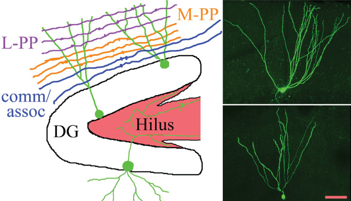
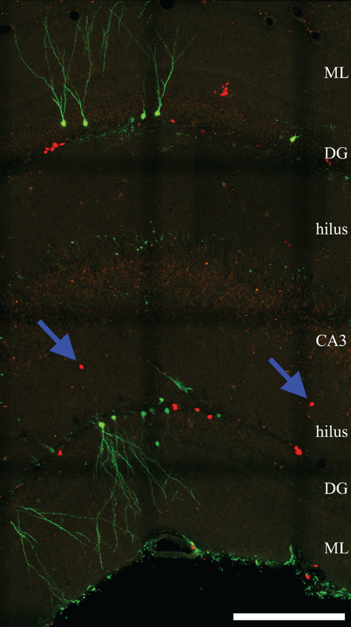
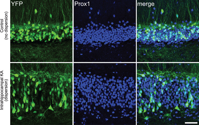
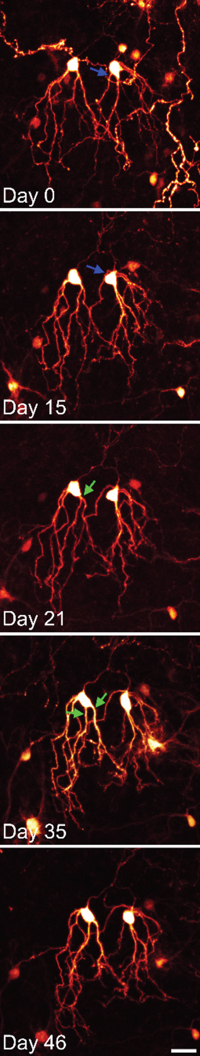
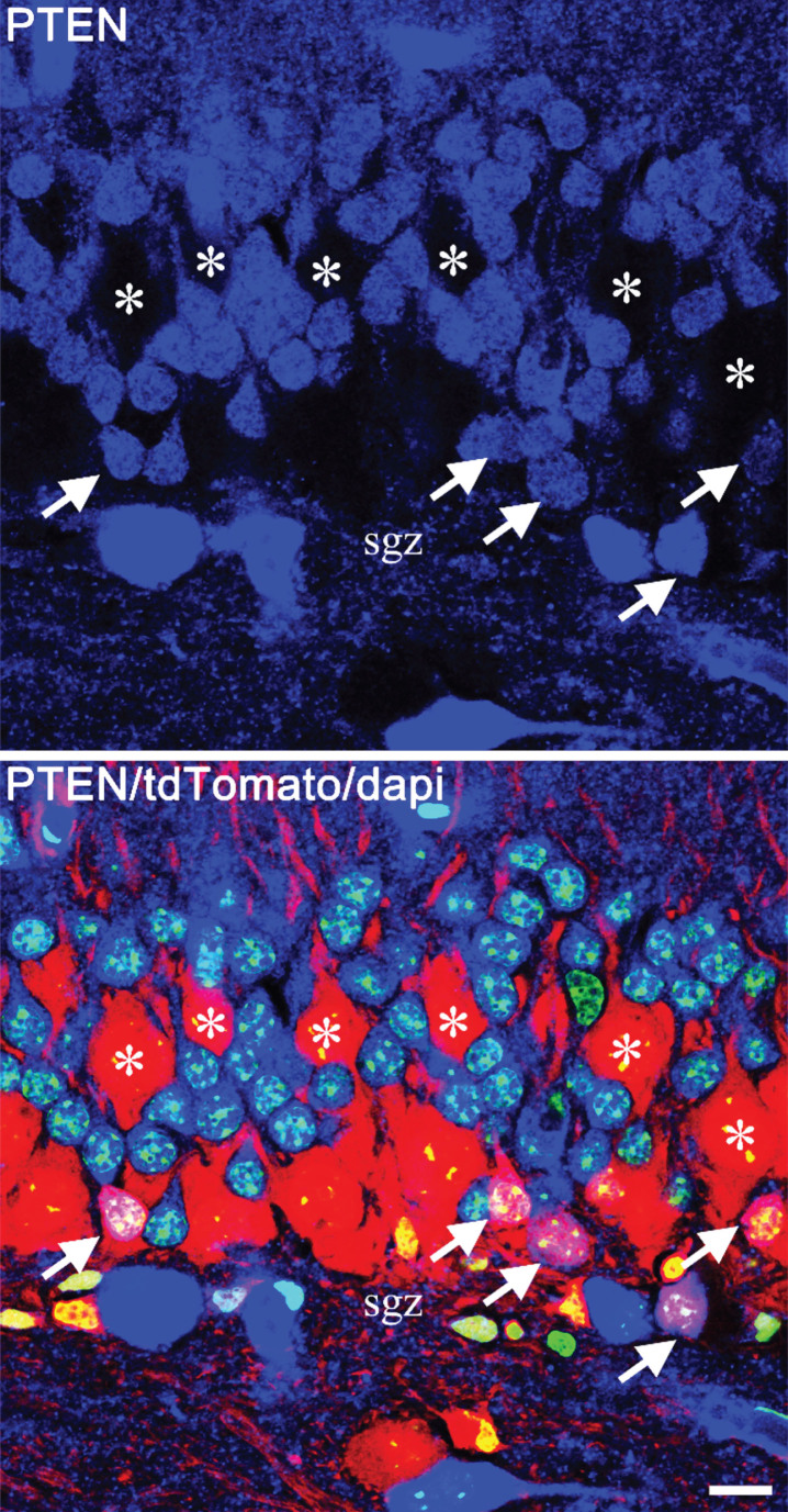
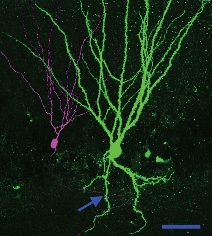
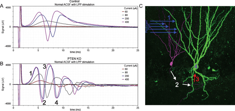
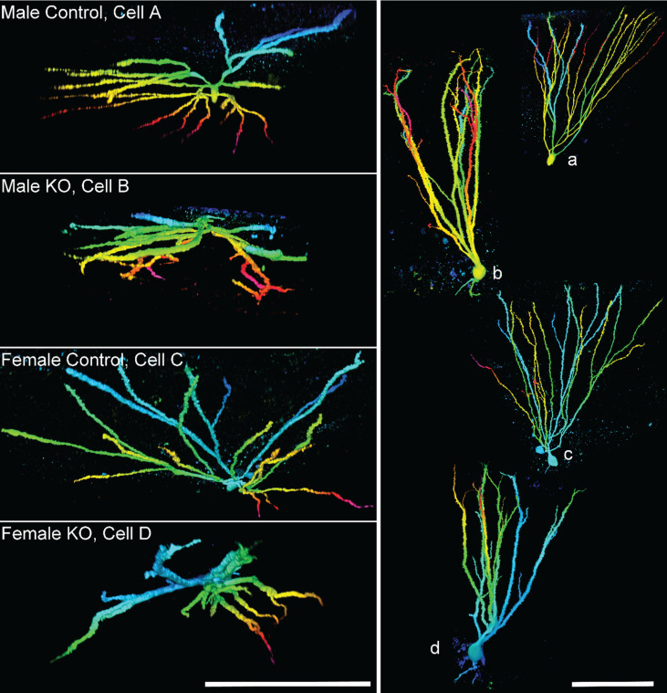
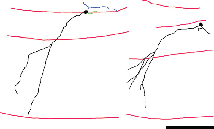
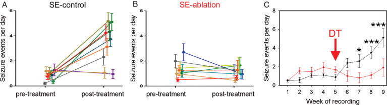
Hippocampal neurogenesis continues throughout life in mammals - including humans. During the development of temporal lobe epilepsy, newly-generated hippocampal granule cells integrate abnormally into the brain. Abnormalities include ectopic localization of newborn cells, de novo formation of abnormal basal dendrites, and disruptions of the apical dendritic tree. Changes in granule cell position and dendritic structure fundamentally alter the types of inputs these cells are able to receive, as well as the relative proportions of remaining inputs. Dendritic abnormalities also create new pathways for recurrent excitation in the hippocampus. These abnormalities are hypothesized to contribute to the development of epilepsy, and may underlie cognitive disorders associated with the disease as well. To test this hypothesis, investigators have used pharmacological and genetic strategies in animal models to alter neurogenesis rates, or ablate the newborn cells outright. While findings are mixed and many unanswered questions remain, numerous studies now demonstrate that ablating newborn granule cells can have disease modifying effects in epilepsy. Taken together, findings provide a strong rationale for continued work to elucidate the role of newborn granule cells in epilepsy: both to understand basic mechanisms underlying the disease, and as a potential novel therapy for epilepsy.

Keywords: Epilepsy; PTEN; basal dendrite; dentate gyrus; mTOR.


Figures

Fig.1
Fig.1
The schematic shows the gross organization of afferent inputs to the molecular layer of the dentate gyrus. Inputs from lateral perforant path (L-PP), medial perforant path (M-PP) and from associational/commissural fibers (comm/assoc) target the outer, middle and inner molecular layer, respectively. Hippocampal granule cells (green) possess cell bodies located in the dentate granule cell body layer (DG) and project dendrites into the molecular layer. Granule cell mossy fiber axons project into the hilus. Micrographs show two biocytin-filled granule cells, revealing the characteristic fanlike spread of the dendritic trees. Scale bar = 50μm.
Fig.2
Fig.2
Confocal maximum projection of YFP- and RFP-expressing newly-generated granule cells from an epileptic Gli1-CreERT2, Gt(ROSA)26Sortm1 (CAG - Brainbow2.1) Cle/J “brainbow” reporter mouse (Singh et al., 2016). Blue arrows denote ectopic RFP-expressing cells located in the dentate hilus. ML, molecular layer. DG, dentate granule cell layer. CA3, CA3 pyramidal cell layer. Scale bar = 200μm.
Fig.3
Fig.3
Confocal images showing granule cell dispersion in the intrahippocampal kainic acid model of epilepsy. Images show YFP-expressing granule cells (green) in the dentate gyrus of a Thy1-YFP transgenic mouse. The Thy1 promoter leads to YFP expression in a subset of CNS neurons, including granule cells. Granule cells are co-labeled with the granule cell marker Prox1. The bottom row shows the dentate gyrus in the hemisphere that was injected with kainic acid, while the top row shows the dentate in the non-injected hemisphere. Pronounced granule cell dispersion and somatic hypertrophy is evident in the injected hemisphere, affecting essentially all granule cells. Scale bar = 50μm.
Fig.4
Fig.4
Time series confocal images of YFP-expressing granule cells in an organotypic hippocampal slice culture. The two granule cells shown were imaged over a period of 6 weeks. Both cells show modest somatic translocation, with the soma moving into the apical dendrites. In both cases, this distorts the apical dendritic tree. The blue arrow shows an apical dendrite, which is shifted to the basal pole of the cell. The green arrows show the soma absorbing a dendritic branch point, converting a single primary dendrite into two primary dendrites. Scale bar = 20μm.
Fig.5
Fig.5
Confocal images showing mosaic deletion of PTEN from newborn hippocampal granule cells in a Gli1-CreERT2, PTENfl/fl, tdTomato reporter mouse. PTEN is shown in blue, tdTomato in red and dapi in green. The top panel shows PTEN alone, and the lower panel shows the merged image. PTEN-expressing newborn cells (arrows) are located in the inner third of the granule cell body layer, close to the subgranular zone (sgz). The much larger PTEN KO granule cells (asterisks), on the other hand, are distributed through the cell body layer, with some migrating close to the molecular layer border. Scale bar = 10μm.
Fig.6
Fig.6
Basal dendrites create a new pathway for recurrent excitatory contacts in the epileptic dentate gyrus. Neuronal reconstruction shows a biocytin-filled PTEN KO cell (green) and a biocytin-filled PTEN-expressing (purple) cell from a PTEN KO (Gli1-CreERT2, PTENfl/fl) mouse. The KO cell exhibits a large basal dendrite. The arrow denotes a point where the axon of the wildtype cell occupies the same focal plane in the z-axis as the PTEN KO cell basal dendrite, suggesting that a synaptic contact could be formed. Scale bar = 50μm.
Fig.7
Fig.7
Responses to lateral perforant path (LPP) stimulation of increasing amplitude (60, 80, 200 and 400μA) from a control mouse and a PTEN KO mouse. In slices from the control mouse (A) the field excitatory post-synaptic potential (fEPSP) increased in amplitude with greater stimulation current and was followed by the appearance of a single population spike (negative going event) once threshold was reached. The slice from the PTEN KO mouse (B) also showed increasing fEPSP slope with increasing current, however, multiple population spikes were evoked. C: Hypothesized mechanism for the generation of multiple population spikes. Perforant path stimulation evokes an fEPSP in granule cell dendrites (1) leading to a population spike (2) which creates a secondary fEPSP in a granule cell basal dendrite (3). This recurrent activation provokes a secondary population spike (4). Portions of this image are reprinted from LaSarge et al. [54].
Fig.8
Fig.8
Images show granule cell reconstructions of PTEN expressing (control) and PTEN knockout (KO) cells from Gli1-CreERT2, PTENfl/fl mice. Cell morphology was revealed by biocytin filling. Cells are projected from above (left, cells A–D), looking down from the top of the dendritic tree towards the soma, and in profile (right, a–d). Note the more limited spread of the dendritic tree among KO cells, and frequent overlapping dendrites. Reconstructions are color-coded by depth. Scale bars = 100μm. Imaged reproduced from Santos et al. [56].
Fig.9
Fig.9
Neurolucida reconstructions of Thy1-GFP-expressing newborn hippocampal granule cells from mice rendered epileptic using the pilocarpine status epilepticus model. Dendrites and somas are shown in black, axons in blue and basal dendrites in green. Red lines denote the hilar—granule cell body layer border, the granule cell body layer—molecular layer border, and the hippocampal fissure, from top to bottom, respectively. The primary dendrites of these granule cells project obliquely into the molecular layer, rather than directly – as is typical for this cell type. The abnormality gives the cells a “windswept” appearance. Scale bar = 100μm. Portions of this image are reprinted from Santos et al. [55].
Fig.10
Fig.10
Cell ablation treatment blocks epilepsy progression. Nestin-CreERT2, DTrfl/fl mice were generated to induce expression of the diphtheria toxin receptor among newborn granule cells. Epilepsy was induced using the pilocarpine status epilepticus model. Following one month of 24/7 video-EEG monitoring, animals were treated with saline (SE-control) or diphtheria toxin (SE-ablation). Pre-treatment and post-treatment seizure frequencies (A,B) are shown for SE-control mice (left, black) and SE-ablation mice (middle, red). Each line shows the means±SEM for one animal. (C) Average number of seizure events during each week of recording for SE-control (black) and SE-ablation (red) groups (DT was given during week 5, red arrow). Ablation treatment prevented the dramatic increase in seizure frequency evident in SE-control animals. *p < 0.05, ***p < 0.001.

References

    1. Leranth C, Hajszan T. Extrinsic afferent systems to the dentate gyrus. Progress in Brain Research. 2007;163:63–84. 10.1016/s0079-6123(07)63004-0 - DOI - PMC - PubMed
    1. Altman J, Bayer SA. Migration and distribution of two populations of hippocampal granule cell precursors during the perinatal and postnatal periods. The Journal of Comparative Neurology. 1990;301(3):365–81. 10.1002/cne.903010304 - DOI - PubMed
    1. Mathews EA, Morgenstern NA, Piatti VC, Zhao C, Jessberger S, Schinder AF, et al. A distinctive layering pattern of mouse dentate granule cells is generated by developmental and adult neurogenesis. The Journal of Comparative Neurology. 2010;518(22):4479–90. 10.1002/cne.22489 - DOI - PMC - PubMed
    1. Kempermann G, Song H, Gage FH. Neurogenesis in the adult hippocampus. Cold Spring Harbor Perspectives in Biology. 2015;7(9):a018812 10.1101/cshperspect.a018812 - DOI - PMC - PubMed
    1. Buckmaster PS. Mossy Fiber Sprouting in the Dentate Gyrus In: th, Noebels JL, Avoli M, Rogawski MA, Olsen RW, Delgado-Escueta AV, editors. Jasper’s Basic Mechanisms of the Epilepsies. Bethesda (MD): National Center for Biotechnology Information (US) Michael A Rogawski, Antonio V Delgado-Escueta, Jeffrey L Noebels, Massimo Avoli and Richard W Olsen; 2012. - PubMed

LinkOut - more resources