. 2016 Nov 19;100(1):94.
doi: 10.5334/jbr-btr.1230.

Cerebellovascular Disease: Magnetic Resonance Imaging

Affiliations

Cerebellovascular Disease: Magnetic Resonance Imaging

Laurens Jaak De Cocker. J Belg Soc Radiol. .

Abstract

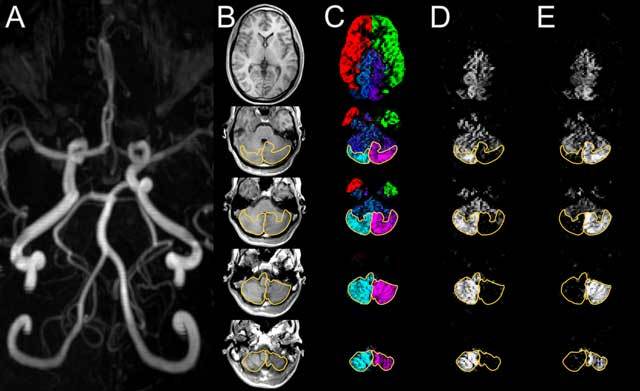
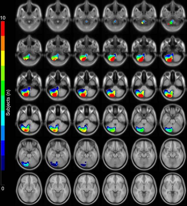
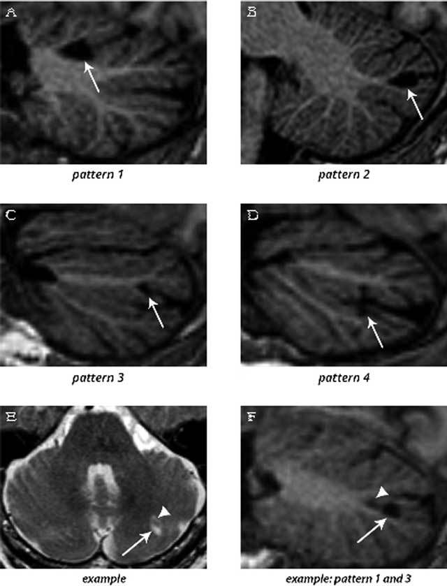
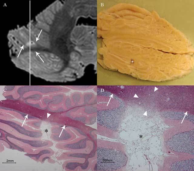
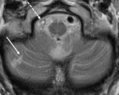
The goal of this thesis was to elucidate the details of cerebellovascular diseases with advanced magnetic resonance (MR) imaging (MRI) and to translate the findings to routine clinical MRI. The first aim was to image cerebellar arterial perfusion territories, which was achieved by applying super-selective arterial spin labelling (ASL) MRI with labelling of both vertebral arteries in addition to the carotid arteries. The second aim was to unravel the imaging patterns of cerebellar infarctions with 7T post-mortem MRI in addition to volume (3D) clinical MRI. This research led to the description of "cerebellar cortical infarct cavities", an incidental imaging finding that proved to be the most frequent manifestation of cerebellar ischemia as well as a marker of atherosclerotic and thromboembolic cerebrovascular disease. Finally, we found that almost all patients with such cavities lack a clinical history of vertebrobasilar transient ischemic attack (TIA) or stroke, disclosing the still very high incidence of clinically occult ischemia in the posterior fossa.

Keywords: 7-Tesla MRI; Arterial Spin Labeling; MRI; PICA; cerebellum; infarct.


Figures

Figure 1
Figure 1
Time-of-flight MRA (A) shows a slightly dominant left vertebral artery. The anatomical T1-weighted images (B) show through the cerebellum from inferior (bottom row) to superior (top row). Territorial perfusion images (C) show the territory supplied by the right (cyan) and left (magenta) vertebral arteries and by the right (red) and left (green) internal carotid arteries. With the mixing of blood in the basilar artery, cyan and magenta turn into blue. Individual perfusion images of the right (D) and left (E) vertebral arteries. The posterior inferior cerebellar artery (PICA) territories are outlined in perfusion images (C–E) and copied to the anatomical images (B). The PICA territories can be easily discriminated; they are symmetrical and supply the posterior cerebellar surfaces (B). Reproduced from Neuroimage [2].
Figure 2
Figure 2
This is a statistical map of the extent of the perfusion territories of the right posterior inferior cerebellar arteries (PICA) as visualized in 10 healthy subjects with super-selective ASL superimposed on a standard MNI space template. The depicted images are orientated axially from inferior (top row) to superior (bottom row) [2].
Figure 3
Figure 3
3D T1-weighted images (sagittal reconstructions) of the brain (A–D and F), providing excellent contrast between gray matter and white matter, show the four patterns by which cerebellar infarcts (arrows) typically affect the cerebellar cortex. An example of two cerebellar infarcts adjacent to each other (E; arrow and arrowhead) are detected on transverse T2WI. Sagittal reconstruction of 3D T1WI of the same two infarcts (F) shows how the topography of one infarct corresponds to pattern 3 (arrow), while the other infarct (arrowhead) corresponds to a small pattern 1 infarct. Notice the sparing of the subcortical and deep white matter in each infarct. The images are cropped to only display the cerebellum. Reproduced from Neuroimage: Clinical [5].
Figure 4
Figure 4
This image shows cerebellar cortical infarct cavity (asterisk) in the right cerebellar hemisphere on post-mortem 7T MRI (A; axial T2WI), gross specimen cut along the line indicated in A (B), and microscopy (C–D; HE stain). Notice the destruction of all three cortical layers on microscopy (C–D) and a preserved juxtacortical white matter (arrows in A, C, and D) with some microscopic gliotic changes (arrowheads in C and D). The surroundings of the cerebellum are dark on T2WI because the specimens were submerged in Fomblin (A), which does not yield MRI signal. Reproduced from Cerebrovascular Diseases [6].
Figure 5
Figure 5
This image shows a small cortical infarct (arrow) in the posterior lobe of the right cerebellar hemisphere related to a thrombosed right vertebral artery (dashed arrow).

References

    1. De Cocker LJ, van Veluw SJ, Fowkes M, Luijten PR, Mali WP, Hendrikse J. Very small cerebellar infarcts: Integration of recent insights into a functional topographic classification. Cerebrovasc Dis. 2013;36:81–87. doi: 10.1159/000353668. - DOI - PubMed
    1. Hartkamp NS, De Cocker LJ, Helle M, et al. In vivo visualization of the PICA perfusion territory with super-selective pseudo-continuous arterial spin labeling MRI. Neuroimage. 2013;83:58–65. doi: 10.1016/j.neuroimage.2013.06.070. - DOI - PubMed
    1. Bokkers RP, De Cocker LJ, van Osch MJP, Hartkamp NS, Hendrikse J. Selective arterial spin labeling: Techniques and neurovascular applications. Top Magn Reson Imaging. 2016;25:73–80. - PubMed
    1. Hartkamp NS, Hendrikse J, De Cocker LJ, de Borst GJ, Kappelle LJ, Bokkers RP. Misinterpretation of ischaemic infarct location in relationship to the cerebrovascular territories [Epub ahead of print] J Neurol Neurosurg Psychiatry. 2016 - PMC - PubMed
    1. De Cocker LJ, Geerlings MI, Hartkamp NS, et al. Cerebellar infarct patterns: The SMART-Medea study. NeuroImage Clin. 2015;8:314–321. doi: 10.1016/j.nicl.2015.02.001. - DOI - PMC - PubMed

LinkOut - more resources