Review
. 2018 Nov;75(22):4093-4105.
doi: 10.1007/s00018-018-2903-z. Epub 2018 Aug 27.

The emerging roles of ribosomal histidyl hydroxylases in cell biology, physiology and disease

Affiliations
Review

The emerging roles of ribosomal histidyl hydroxylases in cell biology, physiology and disease

James R Bundred et al. Cell Mol Life Sci. 2018 Nov.

Abstract

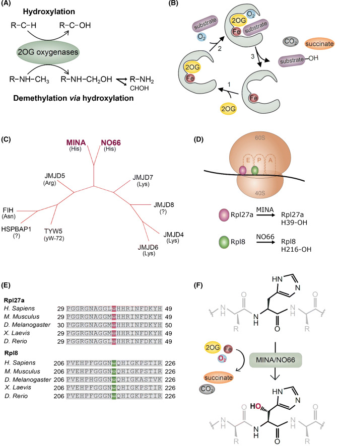
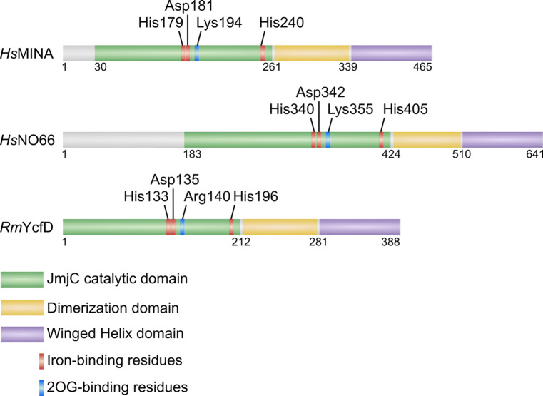
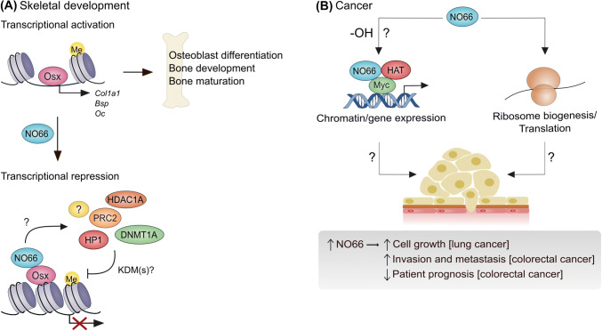
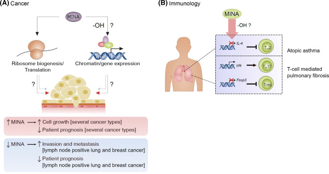
Hydroxylation is a novel protein modification catalyzed by a family of oxygenases that depend on fundamental nutrients and metabolites for activity. Protein hydroxylases have been implicated in a variety of key cellular processes that play important roles in both normal homeostasis and pathogenesis. Here, in this review, we summarize the current literature on a highly conserved sub-family of oxygenases that catalyze protein histidyl hydroxylation. We discuss the evidence supporting the biochemical assignment of these emerging enzymes as ribosomal protein hydroxylases, and provide an overview of their role in immunology, bone development, and cancer.

Keywords: Bone development; Cancer; Histidine; Hydroxylation; Immunology; Post-translational modification.


Conflict of interest statement

The authors report no conflicts of interest.

Figures

Fig. 1
Fig. 1
Ribosomal histidyl hydroxylation catalyzed by the 2OG-oxygenases MINA and NO66. a 2OG-oxygenases that target protein substrates can catalyze stable hydroxylation (top), or demethylation via a hydroxylation reaction (bottom). Demethylation produces the unmethylated substrates and formaldehyde (CHOH). Note that only demethylation of a mono-methylated amino acid is shown: 2OG-oxygenase-mediated demethylation is also possible at tri- and di-methylated residues. b The catalytic cycle of 2OG-oxygenases. For clarity, a graphical representation of only the catalytic pocket, not the whole DSBH domain, is shown. c The JmjC-only family of 2OG-oxygenases. Phylogenetic tree constructed using iTOL online software [80] in unrooted tree format using tree data derived from Clustal Omega alignment of the following human protein sequences; MINA (Q8IUF8), NO66 (Q9H6W3), JMJD4 (Q9H9V9), JMJD5 (Q8N371), JMJD6 (Q6NYC1), JMJD7 (POC870), JMJD8 (Q96S16), TYW5 (A2RUC4), FIH (HIF1AN; Q9NWT6) and HSPBAP1 (Q96EW2). Primary biochemical specificities are indicated by His (histidyl hydroxylase), Lys (lysyl hydroxylase), Arg (arginyl hydroxylase), Asn (asparaginyl hydroxylase), yW-72 (hydroxylase of modified Wybutosine nucleoside in tRNAPhe) or ‘?’ (unknown) [, , , –83]. d Ribosomal oxygenases target important functional domains within the ribosome. MINA targets His-39 of Rpl27a within the large (60S) subunit, which is located close to the ‘E’-site, the binding site of the exiting tRNA. NO66 targets His-216 of the 60S subunit protein Rpl8, which is proximal to the peptidyl transferase centre (PTC). The PTC binds to the P- and A-site tRNAs and catalyzes peptide bond formation. e The histidyl residues hydroxylated by MINA and NO66 are highly conserved. f MINA and NO66 catalyze beta histidyl hydroxylation. NMR studies indicate that hydroxylation occurs on the beta carbon [6]
Fig. 2
Fig. 2
Domain organization of ribosomal oxygenases. Reported crystal structures indicate a unique topology for JmjC ribosomal oxygenases that is conserved from Homo sapiens (Hs) to prokaryotes (including R. marinus, Rm). Note that YcfD catalyzes arginyl hydroxylation of Rpl16 in prokaryotes [6]. Critical catalytic residues are indicated. The dimerization domain is required for oligomerization and activity [45]. The function of the WH domain is unclear but, because of its overall negative charge, is thought unlikely to mediate an interaction with nucleic acid [45]
Fig. 3
Fig. 3
NO66 is implicated in skeletal development and tumorigenesis. a NO66 represses the osteoblast-specific transcription factor Osterix (Osx). Osterix drives the transcription of specific target genes (e.g., Col1a1) to promote osteoblast differentiation and bone development. The dimerization domain of NO66 interacts with the transactivation domain of Osx, leading to reduced Osx target gene expression. Whether this involves a direct effect of an NO66 histone demethylase activity, or results from the recruitment of multiple epigenetic modifiers (e.g., PRC2, HP1, etc.), is currently unclear (signified by ‘?’). b NO66 is over-expressed in lung and colorectal cancers and promotes tumor cell growth and invasion in vitro. Whether the enzymatic activity of NO66 is involved is unclear (‘-OH?’). The molecular mechanisms involved are also unknown, but could be related to roles in myc-driven transcriptional control (left) and/or ribosome biogenesis/translation (right). HDAC1A histone deacetylase 1A; PRC2 polycomb repressor complex 2; HP1 heterochromatin protein 1; DNMT1A DNA methyltransferase 1A; Col1a1 collagen type I alpha 1 chain; Bsp bone sialoprotein; Oc osteocalcin; HAT histone acetyltransferase complex (TRRAP and TIP60)
Fig. 4
Fig. 4
MINA is implicated in tumorigenesis and T-cell differentiation. a MINA is over-expressed in multiple tumor types and is required for cancer cell proliferation in vitro. Whether the enzymatic activity of MINA is required is unclear, particularly with respect to potential roles in gene expression control (‘-OH’?). ‘Driver’ roles for MINA in cancer are denoted by red arrows. MINA may also have tumor suppressor activity in some contexts, possibly via regulation of invasion/metastasis (denoted by blue ‘flat head’ arrows). These paradoxical effects were first uncovered by pathology analyses in lung and breast cancer (summarized at the bottom). b Immunology: MINA regulates the differentiation of specific T-cell subsets in asthma and pulmonary fibrosis. MINA represses Interleukin-4 (IL-4) transcription to regulate T-helper 2 (TH2) cell bias (top). MINA upregulates a T-helper 17 (TH17) differentiation transcriptional program and suppresses the expression of the master regulator of regulatory T-cell (Treg) differentiation (Foxp3)

References

    1. Walsh CT, Garneau-Tsodikova S, Gatto GJ. Protein posttranslational modifications: the chemistry of proteome diversifications. Angew Chem Int Ed. 2005;44(45):7342–7372. doi: 10.1002/anie.200501023. - DOI - PubMed
    1. Markolovic S, Wilkins SE, Schofield CJ. Protein hydroxylation catalyzed by 2-Oxoglutarate-dependent oxygenases. J Biol Chem. 2015;290(34):20712–20722. doi: 10.1074/jbc.R115.662627. - DOI - PMC - PubMed
    1. Hausinger RP. FeII/alpha-ketoglutarate-dependent hydroxylases and related enzymes. Crit Rev Biochem Mol Biol. 2004;39(1):21–68. doi: 10.1080/10409230490440541. - DOI - PubMed
    1. Islam MS, Leissing TM, Chowdhury R, Hopkinson RJ, Schofield CJ. 2-Oxoglutarate-dependent oxygenases. Ann Rev Biochem. 2018;87:585–620. doi: 10.1146/annurev-biochem-061516-044724. - DOI - PubMed
    1. Bottger A, Islam MS, Chowdhury R, Schofield CJ, Wolf A. The oxygenase Jmjd6–a case study in conflicting assignments. Biochem J. 2015;468(2):191–202. doi: 10.1042/BJ20150278. - DOI - PubMed

MeSH terms

LinkOut - more resources