. 2019 Jan;76(1):129-145.
doi: 10.1007/s00018-018-2912-y. Epub 2018 Aug 27.

GADD45α alleviates acetaminophen-induced hepatotoxicity by promoting AMPK activation

Affiliations

GADD45α alleviates acetaminophen-induced hepatotoxicity by promoting AMPK activation

Chunmin Li et al. Cell Mol Life Sci. 2019 Jan.

Abstract

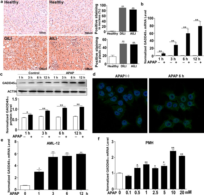
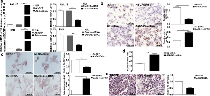
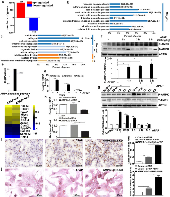
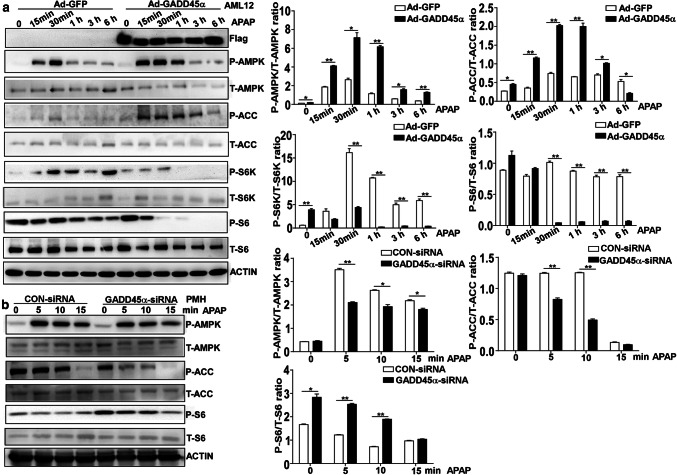
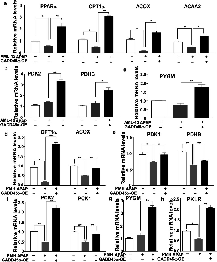
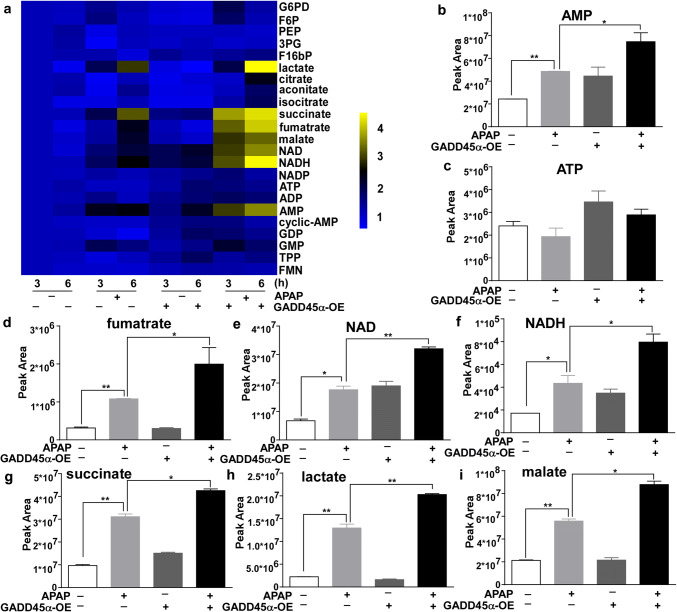
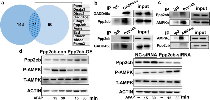
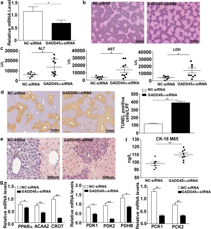
As an analgesic and antipyretic drug, acetaminophen (APAP) is commonly used and known to be safe at therapeutic doses. In many countries, the overuse of APAP provokes acute liver injury and even liver failure. APAP-induced liver injury (AILI) is the most used experimental model of drug-induced liver injury (DILI). Here, we have demonstrated elevated levels of growth arrest and DNA damage-inducible 45α (GADD45α) in the livers of patients with DILI/AILI, in APAP-injured mouse livers and in APAP-treated hepatocytes. GADD45α exhibited a protective effect against APAP-induced liver injury and alleviated the accumulation of small lipid droplets in vitro and in vivo. We found that GADD45α promoted the activation of AMP-activated protein kinase α and induced fatty acid beta-oxidation, tricarboxylic acid cycle (TCA) and glycogenolysis-related gene expression after APAP exposure. Liquid chromatography-mass spectrometry (LC-MS) analysis showed that GADD45α increased the levels of TCA cycle metabolites. Co-immunoprecipitation analysis showed that Ppp2cb, a catalytic subunit of protein phosphatase 2A, could interact directly with GADD45α. Our results indicate that hepatocyte GADD45α might represent a therapeutic target to prevent and rescue liver injury caused by APAP.

Keywords: AMPK; Acetaminophen (APAP); Drug-induced liver injury (DILI); GADD45α.


Figures

Fig. 1
Fig. 1
GADD45α is upregulated during the process of APAP-induced liver injury. a GADD45α expression in the liver was determined by immunohistochemistry and quantified. Human samples from healthy control subjects (Healthy) (n = 3) and from patients with drug-induced liver injury (DILI) (n = 9, four of them APAP-induced DILI). b, c qRT-PCR analysis of the mRNA levels and Western blot analysis of the protein levels of GADD45α in livers from C57BL/6J mice after 300 mg/kg APAP treatment (n = 5 per time point). d Representative images of immunofluorescence staining for GADD45α in AML-12 cells. e qRT-PCR analysis of GADD45α in AML-12 cells after APAP treatment at different time points. f qRT-PCR analysis of GADD45α in primary mouse hepatocytes after different doses of APAP for 6 h. The mRNA levels were normalized to 36B4 and subsequently normalized to the control group. Data are expressed as the mean ± standard error of the mean. *P < 0.05, **P < 0.01
Fig. 2
Fig. 2
GADD45α shows an important capability of protecting against APAP-induced hepatocytes’ death and decreasing the APAP-induced accumulation of small lipid droplets. a The mRNA expression levels of the GADD45α after overexpression and siRNA treatment. b Representative images of TUNEL staining in primary mouse hepatocytes after the overexpression or knockdown of GADD45α at 6 h after APAP treatment and the quantification of the number of TUNEL-positive cells. c Oil Red O staining of primary mouse hepatocytes after the overexpression or knockdown of GADD45α at 6 h after APAP treatment and the quantification of the ratio relative to the control group. d PI staining of AML-12 cells after knockdown of GADD45α at 24 h after APAP treatment. e Oil Red O staining of AML-12 cells from the overexpression of GADD45α at 6 h after APAP treatment and the quantification of the ratio relative to the control group. Data are expressed as the mean ± standard error of the mean. *P < 0.05, **P < 0.01. LPF, low power field
Fig. 3
Fig. 3
The AMPK pathway is activated after exposure to APAP. a Diagram illustrating the genes that were upregulated and downregulated at 6 h. b, c Significantly enriched biological process category in the gene ontology (GO) analysis concerning the genes that were upregulated or downregulated at 6 h after APAP treatment. d The mRNA expression levels of the GADD45 family members (GADD45α, GADD45β and GADD45γ). e Diagram displaying the activation of the AMPK signaling pathway and a heat map representing the log(10) fold change for the genes relating to the AMPK signaling pathway in the APAP treatment group compared with the control group. f Western blot analysis of P-AMPK and T-AMPK in the livers of C57BL/6J mice after treatment with 300 mg/kg APAP for different times. g The protein levels of P-AMPK and T-AMPK in AML-12 cells after APAP treatment for different times. h The mRNA expression levels of AMPKα1 and AMPKα2 in primary mouse hepatocytes after transfections with siRNA targeting AMPKα, confirming the knocking down of AMPKα. (i) TUNEL staining in primary mouse hepatocytes with AMPKα knockdown at 6 h after APAP treatment and the quantification of the number of TUNEL-positive cells. j Oil Red O staining of primary mouse hepatocytes with AMPKα knockdown at 6 h after APAP treatment and the quantification of the ratio relative to the control group. Data are expressed as the mean ± standard error of the mean. *P < 0.05, **P < 0.01
Fig. 4
Fig. 4
GADD45α promotes AMPK activation in APAP-induced hepatocyte injury. a Western blot analysis for P-AMPK, T-AMPK, P-ACC, T-ACC, P-S6K, T-S6K, P-S6 and T-S6 expression in AML-12 cells with overexpression of GADD45α in response to APAP treatment for 0, 0.25, 0.5, 1, 3 and 6 h, followed with quantified protein levels of the above genes. b Western blot analysis for P-AMPK, T-AMPK, P-ACC, T-ACC, P-S6 and T-S6 expression in primary mouse hepatocytes transfected with NC-siRNA or GADD45α-siRNA in response to APAP treatment for 0, 5, 10 and 15 min, followed by quantified protein levels of the above genes. Data are expressed as the mean ± standard error of the mean, *P < 0.05, **P < 0.01
Fig. 5
Fig. 5
Enhanced expression of GADD45α affects fatty acid beta-oxidation-, tricarboxylic acid cycle- and glycogenolysis-related gene expression after APAP treatment. ac The mRNA levels of genes involved in fatty acid beta-oxidation, tricarboxylic acid cycle and glycogenolysis in AML-12 cells with the enhanced expression of GADD45α after APAP treatment for 6 h. dh The mRNA levels of genes involved in fatty acid beta-oxidation, tricarboxylic acid cycle and glycogenolysis in primary mouse hepatocytes with overexpression of GADD45α after APAP treatment for 6 h. The relative mRNA levels were normalized to 36B4 and subsequently normalized to the control group. Data are expressed as the mean ± standard error of the mean, *P < 0.05, **P < 0.01
Fig. 6
Fig. 6
Overexpressed GADD45α can increase the levels of tricarboxylic acid (TCA) cycle metabolites after APAP treatment. a The heat map shows the changes in the levels of 23 metabolites after APAP treatment. bi The relative peak areas of the metabolites involved in the TCA cycle. Data are expressed as the mean ± standard error of the mean. *P < 0.05, **P < 0.01
Fig. 7
Fig. 7
Ppp2cb interacts with GADD45α in hepatocytes. a The diagram represents a total of 11 proteins that were found in two independent co-immunoprecipitation experiments followed by mass spectrometry analysis. b GADD45α (Ppp2cb) was immunoprecipitated from AML-12 cell lysates with enhanced GADD45α expression. Then, the precipitate was evaluated for the presence of Ppp2cb (GADD45α) by Western blot. c AML-12 cells were treated with APAP for 6 h. Then, Ppp2cb (AMPKα) was immunoprecipitated with anti-Ppp2cb (anti-AMPKα), and the presence of AMPKα (Ppp2cb) in the precipitate was evaluated with an anti-AMPKα (anti-Ppp2cb) antibody. d The protein levels of Ppp2cb, P-AMPK and T-AMPK in AML-12 cells were analyzed by Western blot
Fig. 8
Fig. 8
Knockdown of GADD45α aggravated APAP-induced liver injury in vivo. a The mRNA expression levels of the GADD45α after GADD45α-siRNA transfection in mice. b Liver histological examination with H&E; typical images were chosen from each experimental group. c Serum was harvested at 24 h after APAP injection for the measurement of ALT, AST and LDH in the mice of GADD45α-siRNA and control groups. d Representative images of TUNEL staining after the knockdown of GADD45α in mice at 24 h after APAP treatment and the quantification of the number of TUNEL-positive cells. e Oil Red O staining after the knockdown of GADD45α in mice at 24 h after APAP treatment. f Serum levels of CK18 M65, in the GADD45α-siRNA or control groups. g The mRNA levels of genes involved in fatty acid beta-oxidation, tricarboxylic acid cycle and glycogenolysis in the mice with knockdown of GADD45α after APAP treatment for 24 h. The relative mRNA levels were normalized to 36B4 and subsequently normalized to the control group. Data are expressed as the mean ± standard error of the mean, *P < 0.05, **P < 0.01

Similar articles

Cited by

References

    1. Bjornsson ES, Bergmann OM, Bjornsson HK, Kvaran RB, Olafsson S. Incidence, presentation, and outcomes in patients with drug-induced liver injury in the general population of Iceland. Gastroenterology. 2013;144(7):1419–1425. doi: 10.1053/j.gastro.2013.02.006. - DOI - PubMed
    1. Lin CC, Tsai P, Sun HY, Hsu MC, Lee JC, Wu IC, Tsao CW, Chang TT, Young KC. Apolipoprotein J, a glucose-upregulated molecular chaperone, stabilizes core and NS5A to promote infectious hepatitis C virus virion production. J Hepatol. 2014;61(5):984–993. doi: 10.1016/j.jhep.2014.06.026. - DOI - PubMed
    1. Bernal W, Auzinger G, Dhawan A, Wendon J. Acute liver failure. Lancet. 2010;376(9736):190–201. doi: 10.1016/S0140-6736(10)60274-7. - DOI - PubMed
    1. Jaeschke H, McGill MR, Williams CD, Ramachandran A. Current issues with acetaminophen hepatotoxicity—a clinically relevant model to test the efficacy of natural products. Life Sci. 2011;88(17–18):737–745. doi: 10.1016/j.lfs.2011.01.025. - DOI - PMC - PubMed
    1. Masubuchi Y, Suda C, Horie T. Involvement of mitochondrial permeability transition in acetaminophen-induced liver injury in mice. J Hepatol. 2005;42(1):110–116. doi: 10.1016/j.jhep.2004.09.015. - DOI - PubMed

MeSH terms