

Rainer W. Guillery and the genetic analysis of brain development
- PMID: 30152010
- PMCID: PMC6393213
- DOI: 10.1111/ejn.14135
Rainer W. Guillery and the genetic analysis of brain development
Abstract
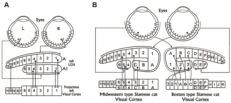
Ray Guillery had broad research interests that spanned cellular neuroanatomy, but was perhaps best known for his investigation of the connectivity and function of the thalamus, especially the visual pathways. His work on the genetics of abnormal vision in albino mammals served as an early paradigm for genetic approaches for studying brain connectivity of complex species in general, and remains of major relevance today. This work, especially on the Siamese cat, illustrates the complex relationship between genotype and physiology of cerebral cortical circuits, and anticipated many of the issues underlying the imperfect relationship between genes, circuits, and behavior in mammalian species including human. This review also briefly summarizes studies from our own lab inspired by Ray Guillery's legacy that continues to explore the relationship between genes, structure, and behavior in human cerebral cortex.
Keywords: Siamese cat; brain development; cerebral cortex; genetics.
© 2018 Federation of European Neuroscience Societies and John Wiley & Sons Ltd.
Figures
Similar articles
-
Conversations with Ray Guillery on albinism: linking Siamese cat visual pathway connectivity to mouse retinal development.Eur J Neurosci. 2019 Apr;49(7):913-927. doi: 10.1111/ejn.14396. Epub 2019 Apr 23. Eur J Neurosci. 2019. PMID: 30801828 Free PMC article. Review.
-
The strengths of the genetic approach to understanding neural systems development and function: Ray Guillery's synthesis.Eur J Neurosci. 2019 Apr;49(7):888-899. doi: 10.1111/ejn.13985. Epub 2018 Aug 1. Eur J Neurosci. 2019. PMID: 29883004 Free PMC article. Review.
-
Development, organization and plasticity of auditory circuits: Lessons from a cherished colleague.Eur J Neurosci. 2019 Apr;49(8):990-1004. doi: 10.1111/ejn.13979. Epub 2018 Aug 16. Eur J Neurosci. 2019. PMID: 29804304 Free PMC article. Review.
-
Neural mechanisms of sensorimotor transformation and action selection.Eur J Neurosci. 2019 Apr;49(8):1055-1060. doi: 10.1111/ejn.14069. Epub 2018 Aug 7. Eur J Neurosci. 2019. PMID: 30019473 Free PMC article. Review.
-
Editorial: Introduction to the special issue in honor of Ray Guillery.Eur J Neurosci. 2019 Apr;49(7):884-887. doi: 10.1111/ejn.14419. Eur J Neurosci. 2019. PMID: 30968456 No abstract available.
Cited by
-
Conversations with Ray Guillery on albinism: linking Siamese cat visual pathway connectivity to mouse retinal development.Eur J Neurosci. 2019 Apr;49(7):913-927. doi: 10.1111/ejn.14396. Epub 2019 Apr 23. Eur J Neurosci. 2019. PMID: 30801828 Free PMC article. Review.
-
Cortical layer with no known function.Eur J Neurosci. 2019 Apr;49(7):957-963. doi: 10.1111/ejn.13978. Epub 2018 Aug 3. Eur J Neurosci. 2019. PMID: 29806977 Free PMC article. No abstract available.
References
-
- Adams NC, Lozsadi DA & Guillery RW (1997) Complexities in the thalamocortical and corticothalamic pathways. Eur J Neurosci, 9, 204–209. - PubMed
-
- Allen KM, Gleeson JG, Shoup SM & Walsh CA (1998) A YAC contig in Xq22.3-q23, from DXS287 to DXS8088, spanning the brain-specific genes doublecortin (DCX) and PAK3. Genomics, 52, 214–218. - PubMed
-
- Bernier R, Golzio C, Xiong B, Stessman HA, Coe BP, Penn O, Witherspoon K, Gerdts J, Baker C, Vulto-van Silfhout AT, Schuurs-Hoeijmakers JH, Fichera M, Bosco P, Buono S, Alberti A, Failla P, Peeters H, Steyaert J, Vissers L, Francescatto L, Mefford HC, Rosenfeld JA, Bakken T, O’Roak BJ, Pawlus M, Moon R, Shendure J, Amaral DG, Lein E, Rankin J, Romano C, de Vries BBA, Katsanis N & Eichler EE (2014) Disruptive CHD8 mutations define a subtype of autism early in development. Cell, 158, 263–276. - PMC - PubMed
-
- Bridge H, von dem Hagen EA, Davies G, Chambers C, Gouws A, Hoffmann M & Morland AB (2014) Changes in brain morphology in albinism reflect reduced visual acuity. Cortex, 56, 64–72. - PubMed
-
- Brodmann K & Garey LJ (2006) Brodmann’s Localisation in the Cerebral Cortex. Springer, New York.
Publication types
MeSH terms
Personal name as subject
- Actions
Grants and funding
LinkOut - more resources
Full Text Sources
Other Literature Sources
Miscellaneous
