Review
. 2018;12(1):284-290.
doi: 10.1080/19336950.2018.1502585.

Cystic fibrosis transmembrane conductance regulator (CFTR): Making an ion channel out of an active transporter structure

Affiliations
Review

Cystic fibrosis transmembrane conductance regulator (CFTR): Making an ion channel out of an active transporter structure

Paul Linsdell. Channels (Austin). 2018.

Abstract

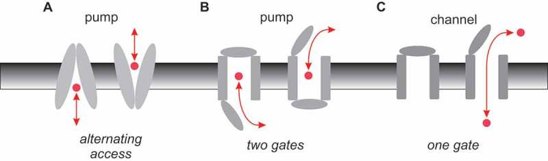
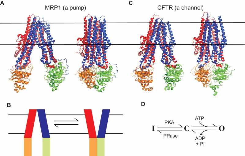
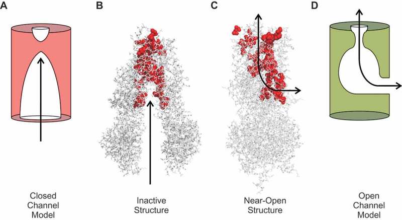
Cystic fibrosis is caused by mutations in the cystic fibrosis transmembrane conductance regulator (CFTR). CFTR is a member of the ATP-binding cassette (ABC) family of membrane transport proteins, most members of which function as ATP-dependent pumps. CFTR is unique among human ABC proteins in functioning not as a pump, but as an ion channel. Recent structural data has indicated that CFTR shares broadly similar overall architecture and ATP-dependent conformational changes as other ABC proteins. Functional investigations suggest that CFTR has a unique open portal connecting the cytoplasm to the transmembrane channel pore, that allows for a continuous pathway for Cl- ions to cross the membrane in one conformation. This lateral portal may be what allows CFTR to function as an ion channel rather than as a pump, suggesting a plausible mechanism by which channel function may have evolved in CFTR.

Keywords: ABC protein; CFTR; channel pore; chloride channel; cystic fibrosis; ion channel.


Figures

Figure 1.
Figure 1.
Basic mechanisms of pump and channel function. (a) Alternating access model of pump function. A conformational change in the membrane-spanning parts of the protein (grey) results in substrate (red) being accessible to the intracellular side of the membrane in one conformation (inward-facing), and to the extracellular side of the membrane in the other conformation (outward-facing). (b) This same mechanism can be expressed in terms of outer and inner gates that are never open at the same time (outer gate closed in the inward-facing state, inner gate closed in the outward-facing state). (c) Ion channels can operate with a single gate, which is closed in the channel closed state and open in the channel open state, allowing a continuous aqueous pathway for ion electrodiffusion across the membrane.
Figure 2.
Figure 2.
Atomic structures of MRP1 and CFTR. (a) Cryo-EM structures of bovine MRP1 in the inward-facing state (NBDs separated; left) [18] and in the outward-facing state (NBDs dimerized; right) [19]. MSD1, red; MSD2, blue; NBD1, orange; NBD2, green. The approximate location of the cell membrane is indicated by horizontal black lines. To facilitate comparison with the structure of CFTR, an additional N-terminal membrane-associated domain found in MRP1 (MSD0) has been removed from these images. (b) These structures are consistent with an NBD-controlled, alternating access mechanism of active transport by MRP1 [19], as proposed for other ABC proteins [4,15,16]. (c) Cryo-EM structures of human CFTR in an inactive, dephosphorylated state (left) [12], and zebrafish CFTR in a “near-open” state that is closed only at the extracellular ends of the MSDs (right) [13]. The MSDs and NBDs are shown in the same colour scheme as in (a). The cytoplasmic R domain, which is mostly unstructured, is not shown in these images. (d) Basic minimal model for CFTR functional regulation by phosphorylation and by ATP. The channel exists in an inactive state (i) until phosphorylation of the R domain by protein kinase A (PKA). Phosphorylated CFTR transitions to the open burst state (o) following ATP binding, and returns to the closed interburst state (c) following ATP hydrolysis and the release of hydrolysis products (ADP and Pi). ATP-dependent gating continues until the R domain is dephosphorylated by phosphoprotein phosphatases (PPase). This minimal model is a gross oversimplification of current understanding of CFTR channel gating [6,24] to emphasize that the structure shown in (b) (left) corresponds to state I, while that on the right is expected to be closest to state O [13]. The structure of the phosphorylated, interburst closed state C, as described in the text, is not currently known.
Figure 3.
Figure 3.
CFTR channel pore-lining residues and location of the cytoplasmic portal. (A-C) Location of putative channel pore-lining amino acid side-chains (red) [23,24] within the CFTR structures shown in Figure 2. (a) In the inactive, dephosphorylated state, a wide central pathway connects the MSDs to the cytoplasm (arrow). (b,c) In the near-open state, this central pathway is closed to the cytoplasm by dimerization of the NBDs and closing of the inner ends of the MSDs. Instead, cytoplasmic access to the pore is via a lateral portal between TMEs 4 and 6. This portal is facing the viewer in (b), and on the right hand side of the MSDs in (c), indicating cytoplasmic access as indicated by the arrows. (d) A cross-section through the TMEs indicates the location of the portal (arrow), as well as important positively charged amino acid side chains (red; K190 (TME3), R248 (TME4), R303 (TME5) and K370 (TME6) in human CFTR) that have been shown to play functional roles in the electrostatic attraction of cytoplasmic anions to the pore [29,30].
Figure 4.
Figure 4.
Proposed mechanisms of CFTR channel opening and closing. (a) Cartoon functional model of the pore in the channel closed state. The pore is wide open to the cytoplasm, but closed close to its extracellular end. (b) This cartoon model is somewhat reminiscent of the inward-facing structure of inactive CFTR (putative pore-lining amino acid side chains shown in red). (c) In the near-open structure, cytoplasmic access to the pore is via a lateral portal. (d) Cartoon functional model of the open channel pore incorporating a single lateral portal on the cytoplasmic side, a relatively wide inner vestibule, and narrow selectivity filter region close to the extracellular end of the pore. See [23] for more details concerning functional models of the open channel pore.

References

    1. Riordan JR, Rommens JM, Kerem B, et al. Identification of the cystic fibrosis gene: cloning and characterization of complementary DNA. Science. 1989;245:1066–1073. - PubMed
    1. Dean M, Rzhetsky A, Alikmets R.. The human ATP-binding cassette (ABC) transporter superfamily. Genome Res. 2001;11:1156–1166. - PubMed
    1. Rees DC, Johnson E, Lewinson O.. ABC transporters: the power to change. Nat Rev Mol Cell Biol. 2009;10:218–227. - PMC - PubMed
    1. Locher KP. Mechanistic diversity in ATP-binding cassette (ABC) transporters. Nat Struct Mol Biol. 2016;23:487–493. - PubMed
    1. Gadsby DC, Vergani P, Csanády L. The ABC protein turned chloride channel whose failure causes cystic fibrosis. Nature. 2006;440:477–483. - PMC - PubMed

MeSH terms

Substances

LinkOut - more resources