. 2018 Nov;18(5):4328-4334.
doi: 10.3892/mmr.2018.9425. Epub 2018 Aug 24.

Protein phosphatase 2A regulates the p38 signaling pathway to affect the migration of astrocytes

Affiliations

Protein phosphatase 2A regulates the p38 signaling pathway to affect the migration of astrocytes

Lijun Zhang et al. Mol Med Rep. 2018 Nov.

Abstract

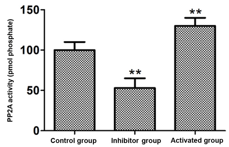
The aim of the present study was to investigate the effect and mechanism of protein phosphatase 2A (PP2A) on the migration of astrocytes. The primary astrocytes of neonatal mice were isolated and cultured in vitro, and treated with the PP2A activator D‑erythro‑sphingosine (DES) (activated group) or inhibitor okadaic acid (inhibitory group). The control group was treated with equal amounts of dimethyl sulfoxide. The activity of PP2A in the cells was detected using a commercial kit and the migration of cells was investigated using a Transwell migration assay. The protein expression of p38, phosphorylated (p)‑p38, matrix metalloproteinase (MMP)‑2 and MMP‑9 was detected by western blotting. Cell migration and the protein expression of p38, p‑p38, MMP‑2 and MMP‑9 was also determined following treatment of astrocytes with the p38 signaling pathway inhibitor SB202190 with or without the PP2A activator DES. The results demonstrated that the activity of PP2A in the PP2A inhibitory group was significantly decreased compared with the control group, while that of the PP2A‑activated cells was significantly increased compared with the control. The protein levels of MMP‑2 and MMP‑9 in the PP2A inhibitory group astrocytes were significantly decreased compared with the control group, while PP2A‑activated astrocytes exhibited significantly increased levels of these proteins. By contrast, the p‑p38 level in PP2A inhibitory group astrocytes was significantly increased compared with the control group, while astrocytes in the activated group exhibited significantly lower levels compared with the control group. Furthermore, the cell migration ability, and MMP‑2 and MMP‑9 protein levels, of astrocytes that received combined treatment with SB202190 and the PP2A activator DES were significantly increased compared with the levels in astrocytes treated with SB202190 alone. The results of the current study indicate that PP2A may negatively regulate the p38 signaling pathway to promote astrocyte migration.


Figures

Figure 1.
Figure 1.
PP2A activity in astrocytes of the control, PP2A inhibitor and PP2A activation groups. **P<0.01 vs. control group. PP2A, protein phosphatase 2A.
Figure 2.
Figure 2.
Effect of PP2A on cell migration and the protein expression of MMPs in astrocytes of the control, PP2A inhibitor and PP2A activation groups. (A) Cell migration was determined by a Transwell migration assay. (B) Representative western blot bands for the protein expression of MMP-2 and MMP-9 in control, PP2A inhibitor and PP2A activation groups. (C) Densitometric analysis was performed to obtain the relative expression level of target proteins with β-actin as the internal reference. **P<0.01 vs. control group. PP2A, protein phosphatase 2A; MMP, matrix metalloproteinase.
Figure 3.
Figure 3.
Effect of PP2A on p-p38 protein expression in astrocytes of the control, PP2A inhibitor and PP2A activation groups. (A) Representative western blot bands for the protein expression of p-p38 and p38 in control, PP2A inhibitor and PP2A activation groups. (B) Densitometric analysis was performed to obtain the relative expression level of p-p38 protein with p38 as the internal reference. **P<0.01 vs. control group. PP2A, protein phosphatase 2A; p-p38, phosphorylated-p38.
Figure 4.
Figure 4.
Effect of a p38 signaling pathway inhibitor on p-p38 protein expression in astrocytes. (A) Representative western blot bands for the protein expression of p-p38 and p38 in untreated astrocytes and astrocytes treated with the p38 signaling pathway inhibitor, SB202190. (B) Densitometric analysis was performed to obtain the relative expression level of p-p38 protein with p38 as the internal reference. **P<0.01 vs. untreated group. p-p38, phosphorylated-p38.
Figure 5.
Figure 5.
Effect of inhibition of the p38 signaling pathway on cell migration and the protein expression of MMPs in astrocytes of the untreated, SB202190 and (DES) + SB202190 groups. (A) Cell migration was determined by a Transwell migration assay. (B) Representative western blot bands for the protein expression of MMP-2 and MMP-9 in the untreated, SB202190 and DES + SB202190 groups. (C) Densitometric analysis was performed to obtain the relative expression level of target proteins with β-actin as the internal reference. SB202190 was employed as an inhibitor of the p38 signaling pathway, while DES was employed as an activator of protein phosphatase 2A. **P<0.01 vs. untreated group; ##P<0.01 vs. SB202190 group. DES, D-erythro-sphingosine; MMP, matrix metalloproteinase.

Similar articles

Cited by

References

    1. Furihata T, Ito R, Kamiichi A, Saito K, Chiba K. Establishment and characterization of a new conditionally immortalized human astrocyte cell line. J Neurochem. 2016;136:92–105. doi: 10.1111/jnc.13358. - DOI - PubMed
    1. Maresca B, Spagnuolo MS, Cigliano L. Haptoglobin modulates beta-amyloid uptake by U-87 MG astrocyte cell line. J Mol Neurosci. 2015;56:35–47. doi: 10.1007/s12031-014-0465-6. - DOI - PubMed
    1. Rajalakshmy AR, Malathi J, Madhavan HN. Serum-derived hepatitis C virus 1a infection of human astrocyte cell line SVG. J Viral Hepat. 2016;23:211–216. doi: 10.1111/jvh.12480. - DOI - PubMed
    1. Ono K, Suzuki H, Higa M, Tabata K, Sawada M. Glutamate release from astrocyte cell-line GL261 via alterations in the intracellular ion environment. J Neural Transm (Vienna) 2014;121:245–257. doi: 10.1007/s00702-013-1096-8. - DOI - PubMed
    1. Liu Y, Zeng X, Hui Y, Zhu C, Wu J, Taylor DH, Ji J, Fan W, Huang Z, Hu J. Activation of α7 nicotinic acetylcholine receptors protects astrocytes against oxidative stress-induced apoptosis: Implications for Parkinson's disease. Neuropharmacology. 2015;91:87–96. doi: 10.1016/j.neuropharm.2014.11.028. - DOI - PubMed

Substances