. 2018 Aug 28;13(8):e0202791.
doi: 10.1371/journal.pone.0202791. eCollection 2018.

OmpA-like proteins of Porphyromonas gingivalis contribute to serum resistance and prevent Toll-like receptor 4-mediated host cell activation

Affiliations

OmpA-like proteins of Porphyromonas gingivalis contribute to serum resistance and prevent Toll-like receptor 4-mediated host cell activation

Megumi Inomata et al. PLoS One. .

Abstract

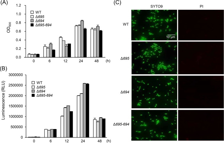
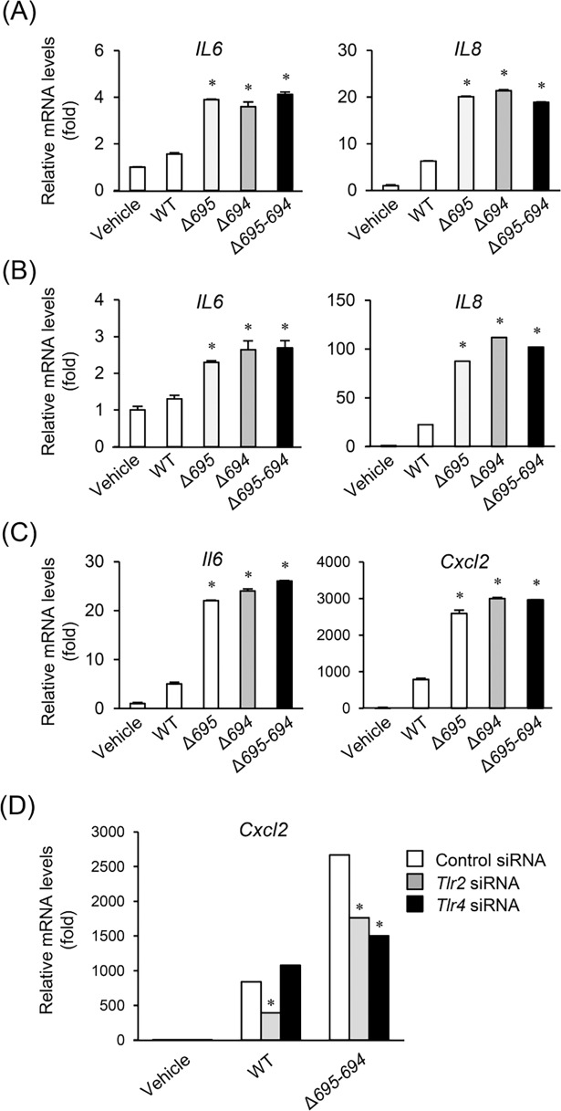
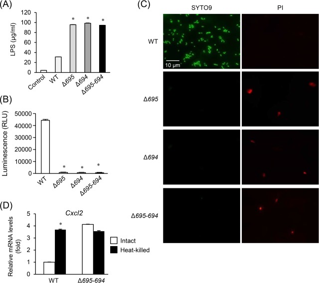
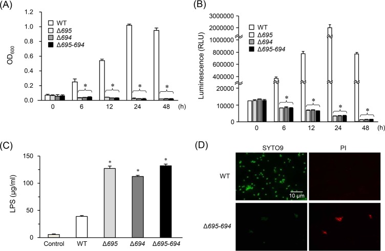
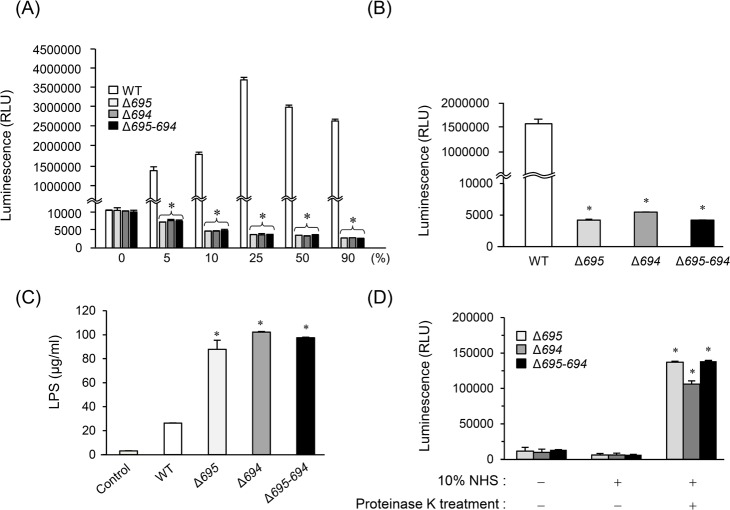
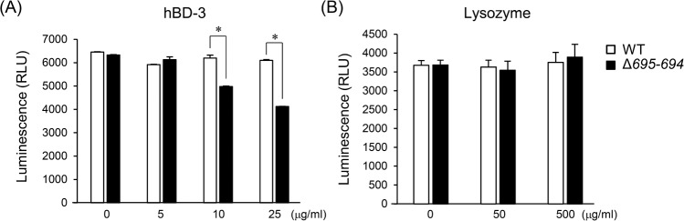
Porphyromonas gingivalis possesses various abilities to evade and disrupt host immune responses, by which it acts as an important periodontal pathogen. P. gingivalis produces outer membrane protein A (OmpA)-like proteins (OmpALPs), Pgm6 and Pgm7, as major O-linked glycoproteins, but their pathological roles in P. gingivalis infection are largely unknown. Here, we report that OmpALP-deficient strains of P. gingivalis show an enhanced stimulatory activity in coculture with host cells. Such an altered ability of the OmpALP-deficient strains was found to be due to their impaired survival in coculture and the release of LPS from dead bacterial cells to stimulate Toll-like receptor 4 (TLR4). Further analyses revealed that the OmpALP-deficient strains were inviable in serum-containing media although they grew normally in the bacterial medium. The wild-type strain was able to grow in 90% normal human serum, while the OmpALP-deficient strains did not survive even at 5%. The OmpALP-deficient strains did not survive in heat-inactivated serum, but they gained the ability to survive and grow in proteinase K-treated serum. Of note, the sensitivity of the OmpALP-deficient strains to the bactericidal activity of human β-defensin 3 was increased as compared with the WT. Thus, this study suggests that OmpALPs Pgm6 and Pgm7 are important for serum resistance of P. gingivalis. These proteins prevent bacterial cell destruction by serum and innate immune recognition by TLR4; this way, P. gingivalis may adeptly colonize serum-containing gingival crevicular fluids and subgingival environments.


Conflict of interest statement

The authors have declared that no competing interests exist.

Figures

Fig 1
Fig 1. Survival and growth of the OmpALP-deficient strains of P. gingivalis in sTSB.
Bacterial cells (107) of the strains WT, Δ695, Δ694, and Δ695–694 were anaerobically cultured in 1 ml of sTSB for the indicated periods. (A and B) The growth was monitored by measuring OD600 (A), and the survival was assessed by ATP production (B). Each value is expressed as mean ± SD (n = 3). (C) The integrity of outer membranes was assessed by double fluorescent staining of bacteria (cultured for 12 h) with SYTO9 and PI. Images were captured by means of a fluorescence microscope.
Fig 2
Fig 2. Involvement of TLR2 and TLR4 in the cell-stimulatory activities of the OmpALP-deficient strain of P. gingivalis.
(A-C) HGFs, HUVECs, and RAW264.7 cells (105) were grown in 12-well plates in an antibiotic-free culture medium containing 10% of inactivated FBS. Bacterial cells of WT, Δ695, Δ694, or Δ695–694 resuspended in PBS were added to the cell cultures (at MOI: 100 for HGFs and HUVECs; at MOI: 10 for RAW264.7 cells). The cells were incubated for 12 h, followed by RNA extraction. Relative expression levels of IL6 and IL8 in HGFs (A) and HUVECs (B) and, Il6 and Cxcl2 in RAW264.7 cells (C) were determined by qRT-PCR. Each value, expressed as a fold increase, is mean ± SD (n = 3); *p < 0.05 (compared to vehicle), one-way ANOVA and Dunnett’s test for post hoc comparisons (μc < μi). (D) RAW264.7 cells were transfected with siRNA targeting Tlr2 or Tlr4 or with control siRNA. Cells cultured in an antibiotic-free culture medium containing 10% of inactivated FBS were then stimulated with bacterial cells of WT or Δ695–694 resuspended in PBS at MOI: 10. The cells were incubated for 12 h, followed by RNA extraction. Relative expression levels of Cxcl2 were determined by qRT-PCR. Each value, expressed as a fold increase, is mean ± SD (n = 3); *p < 0.05 (compared to the Control siRNA), one-way ANOVA and Dunnett’s test for post hoc comparisons (μc ≠ μi).
Fig 3
Fig 3. Survival and growth of the OmpALP-deficient strains of P. gingivalis in DMEM containing 10% of inactivated FBS.
(A–C) Bacterial cells (107) of strains WT, Δ695, Δ694, and Δ695–694 resuspended in PBS were anaerobically cultured in 1 ml of DMEM containing 10% of inactivated FBS for 6 h. LPS concentrations in the culture supernatants were determined (A). Each value is expressed as the mean ± SD (n = 3); *p < 0.05 (compared to PBS control), one-way ANOVA and Dunnett’s test for post hoc comparisons (μc < μi). Their survival was assessed by ATP production (B). Each value is expressed as mean ± SD (n = 3); *p < 0.05 (compared to the WT), one-way ANOVA and Dunnett’s test for post hoc comparisons (μc ≠ μi). The integrity of outer membranes was also assessed by fluorescent staining of bacteria (cultured for 12 h) with SYTO9 and PI (C). Images were captured by means of a fluorescence microscope. (D) Bacterial cells (107) of strains WT and Δ695–694 resuspended in PBS were left untreated (‘Intact’) or boiled for 30 min (‘Heat-killed’). RAW264.7 cells grown in 24-well plates were stimulated with these bacterial cells at MOI 10 for 12 h, followed by RNA extraction. Relative expression levels of Cxcl2 were determined by qRT-PCR. Each value, expressed as a fold increase, is mean ± SD (n = 3); *p < 0.05 (compared to group Intact), one-way ANOVA and Dunnett’s test for post hoc comparisons (μc ≠ μi).
Fig 4
Fig 4. Survival and growth of the OmpALP-deficient strains of P. gingivalis in 10% NHS.
Bacterial cells (107) of strains WT, Δ695, Δ694, and Δ695–694 resuspended in 0.9 ml of PBS were mixed with 0.1 ml of NHS and anaerobically cultured for the indicated periods. (A and B) Their growth was monitored by measuring OD600 (A), and their survival was assessed by ATP production (B). Each value is expressed as mean ± SD (n = 3); *p < 0.05 (compared to the WT), one-way ANOVA and Dunnett’s test for post hoc comparisons (μc ≠ μi). (C) LPS concentrations in the supernatants of these cultures (6 h) were determined (A). Each value is expressed as mean ± SD (n = 3); *p < 0.05 (compared to the Control), one-way ANOVA and Dunnett’s test for post hoc comparisons (μc < μi). (D) The integrity of outer membranes was also assessed by fluorescent staining of bacteria (cultured for 12 h) with SYTO9 and PI. The images were acquired by means of a fluorescence microscope. The results on strains Δ695 and Δ694 are shown in S5 Fig.
Fig 5
Fig 5. Survival and growth of the OmpALP-deficient strains of P. gingivalis in NHS.
(A) Bacterial cells (107) of strains WT, Δ695, Δ694, and Δ695–694 resuspended in 0.1 ml of PBS were mixed with 0.9 ml of indicated concentrations of diluted NHS and anaerobically cultured for 24 h. Bacterial survival was assessed by ATP production. Each value is expressed as mean ± SD (n = 3); *p < 0.05 (compared to the WT), one-way ANOVA and Dunnett’s test for post hoc comparisons (μc ≠ μi). (B and C) Effects of heat-inactivation of NHS. Bacterial cells (107) of strains WT, Δ695, Δ694, and Δ695–694 were cultured in 10% heat-inactivated NHS for 24 h. Bacterial survival was assessed by ATP production (B). LPS concentrations in the supernatants of the cultures (6 h) were determined (C). Each value is expressed as mean ± SD (n = 3); *p < 0.05 (compared to the Control), one-way ANOVA and Dunnett’s test for post hoc comparisons (μc ≠ μi). (D) Effects of proteinase K treatment of NHS. Bacterial cells (107) of strains Δ695, Δ694, and Δ695–694 were cultured in 10% proteinase K-treated or untreated NHS for 24 h. Bacterial survival was assessed by ATP production. Each value is expressed as mean ± SD (n = 3); *p < 0.05, one-way ANOVA and multiple Dunnett’s test for post hoc comparisons between the groups of interest (μc ≠ μi).
Fig 6
Fig 6. The sensitivity of the OmpALP-deficient strains of P. gingivalis to the bactericidal activities of hBD-3 and lysozyme.
(A and B) Bacterial cells (107) of strains WT and Δ695–694, resuspended in 0.25 ml sTSB, were mixed with 0.75 ml of the indicated concentrations of diluted anti-microbial agents and anaerobically cultured for 6 h. Bacterial survival was assessed by ATP production. Each value is expressed as mean ± SD (n = 3); *p < 0.05, one-way ANOVA and Dunnett’s test for post hoc comparisons (μc ≠ μi).

Similar articles

Cited by

References

    1. Lamont RJ, Jenkinson HF. Subgingival colonization by Porphyromonas gingivalis. Oral Microbiol Immunol. 2000;15(6):341–9. - PubMed
    1. Brown LJ, Loe H. Prevalence, extent, severity and progression of periodontal disease. Periodontol 2000. 1993;2:57–71. - PubMed
    1. Ximenez-Fyvie LA, Haffajee AD, Socransky SS. Comparison of the microbiota of supra- and subgingival plaque in health and periodontitis. J Clin Periodontol. 2000;27(9):648–57. - PubMed
    1. Genco R, Offenbacher S, Beck J. Periodontal disease and cardiovascular disease: epidemiology and possible mechanism s. J Am Dent Assoc. 2002;133 Suppl:14S–22S. - PubMed
    1. Scannapieco FA, Genco RJ. Association of periodontal infections with atherosclerotic and pulmonary diseases. J Periodontal Res. 1999;34(7):340–5. - PubMed

Publication types

MeSH terms

LinkOut - more resources