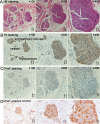
Selective ablation of the ligament of Marshall reduces ischemia and reperfusion-induced ventricular arrhythmias
- PMID: 30153281
- PMCID: PMC6112646
- DOI: 10.1371/journal.pone.0203083
Selective ablation of the ligament of Marshall reduces ischemia and reperfusion-induced ventricular arrhythmias
Abstract
Cardiac sympathetic tone overdrive is a key mechanism of arrhythmia. Cardiac sympathetic nerves denervation, such as LSG ablation or renal sympathetic denervation, suppressed both the prevalence of VAs and the incidence of SCD. Accumulating evidence demonstrates the ligament of Marshall (LOM) is a key component of the sympathetic conduit between the left stellate ganglion (LSG) and the ventricles. The present study aimed to investigate the roles of the distal segment of LOM (LOMLSPV) denervation in ischemia and reperfusion (IR)-induced VAs, and compared that LSG denervation. Thirty-three canines were randomly divided into group 1 (IR group, n = 11), group 2 (LOMLSPV Denervation + IR, n = 9), and group 3 (LSG Denervation + IR, n = 13). Hematoxylin-Eosin (HE) and Immunohistochemistry staining revealed that LOMLSPV contained bundles of sympathetic but not parasympathetic nerves. IR increased the cardiac sympathetic tone [serum concentrations of noradrenaline (NE) and epinephrine (E)] and induced the prevalence of VAs [ventricular premature beat (VPB), salvo of VPB, ventricular tachycardia (VT), VT duration (VTD) and ventricular fibrillation (VF)]. Both LOMLSPV denervation and LSG denervation could reduce the cardiac sympathetic tone in Baseline (BS) [heart rate variability (HRV)]. Compared with group 1, LOMLSPV denervation and LSG denervation similarly reduced sympathetic tone [NE (1.39±0.068 ng/ml in group 2, 1.29±0.081 ng/ml in group 3 vs 2.32±0.17 ng/ml in group 1, P<0.05) and E (114.64±9.22 pg/ml in group 2, 112.60±9.69 pg/ml in group 3 vs 166.18±15.78 pg/ml in group 1, P<0.05),] and VAs [VT (0±3.00 in group 2, 0±1.75 in group 3 vs 8.00±11.00 in group 1, P<0.05) and VTD (0 ± 4 s in group 2, 0±0.88s in group 3 vs 10.0 ± 22.00s in group 1, P<0.05)] after 2h reperfusion. These findings indicated LOMLSPV denervation reduced the prevalence of VT by suppressing SNS activity. These effects are comparable to those of LSG denervation. In myocardial IR, the anti-arrhythmic effects of LOMLSPV Denervation may be related to the inhibition of the expression of NE and E.
Conflict of interest statement
The authors have declared that no competing interests exist.
Figures


Similar articles
-
Ablation of the Ligament of Marshall and Left Stellate Ganglion Similarly Reduces Ventricular Arrhythmias During Acute Myocardial Infarction.Circ Arrhythm Electrophysiol. 2018 May;11(5):e005945. doi: 10.1161/CIRCEP.117.005945. Circ Arrhythm Electrophysiol. 2018. PMID: 29700056
-
Selective Ablation of the Ligament of Marshall Reduces the Prevalence of Ventricular Arrhythmias Through Autonomic Modulation in a Cesium-Induced Long QT Canine Model.JACC Clin Electrophysiol. 2016 Feb;2(1):97-106. doi: 10.1016/j.jacep.2015.09.014. Epub 2015 Nov 10. JACC Clin Electrophysiol. 2016. PMID: 29766860
-
Selective ablation of ligament of Marshall inhibits ventricular arrhythmias during acute myocardial infarction: Possible mechanisms.J Cardiovasc Electrophysiol. 2019 Mar;30(3):374-382. doi: 10.1111/jce.13802. Epub 2018 Dec 19. J Cardiovasc Electrophysiol. 2019. PMID: 30516302
-
Denervation of the extrinsic cardiac sympathetic nervous system as a treatment modality for arrhythmia.Europace. 2017 Jul 1;19(7):1075-1083. doi: 10.1093/europace/eux011. Europace. 2017. PMID: 28340164 Review.
-
Cardiac Sympathetic Denervation for the Management of Ventricular Arrhythmias.J Interv Card Electrophysiol. 2022 Dec;65(3):813-826. doi: 10.1007/s10840-022-01211-2. Epub 2022 Apr 9. J Interv Card Electrophysiol. 2022. PMID: 35397706 Review.
Cited by
-
Vagus Nerve Stimulation by Focused Ultrasound Attenuates Acute Myocardial Ischemia/Reperfusion Injury Predominantly Through Cholinergic Anti-inflammatory Pathway.Cardiovasc Drugs Ther. 2025 Jun 2. doi: 10.1007/s10557-025-07718-w. Online ahead of print. Cardiovasc Drugs Ther. 2025. PMID: 40455110
-
The protective role of vagus nerve stimulation in ischemia-reperfusion injury.Heliyon. 2024 May 9;10(10):e30952. doi: 10.1016/j.heliyon.2024.e30952. eCollection 2024 May 30. Heliyon. 2024. PMID: 38770302 Free PMC article. Review.
-
Autonomic Neuromodulation for Preventing and Treating Ventricular Arrhythmias.Front Physiol. 2019 Mar 11;10:200. doi: 10.3389/fphys.2019.00200. eCollection 2019. Front Physiol. 2019. PMID: 30914967 Free PMC article. Review.
References
-
- Wang S, Zhou X, Huang B, Wang Z, Liao K, Saren G, et al. Spinal cord stimulation protects against ventricular arrhythmias by suppressing left stellate ganglion neural activity in an acute myocardial infarction canine model. Heart Rhythm. 2015; 12: 1628–1635. 10.1016/j.hrthm.2015.03.023 - DOI - PubMed
-
- Schneider HE, Steinmetz M, Krause U, Kriebel T, Ruschewski W, Paul T. Left cardiac sympathetic denervation for the management of life-threatening ventricular tachyarrhythmias in young patients with catecholaminergic polymorphic ventricular tachycardia and long QT syndrome. Clin Res Cardiol. 2013; 102: 33–42. 10.1007/s00392-012-0492-7 - DOI - PMC - PubMed
Publication types
MeSH terms
Substances
LinkOut - more resources
Full Text Sources
Other Literature Sources
Medical

