. 2018 Nov:182:312-322.
doi: 10.1016/j.biomaterials.2018.08.027. Epub 2018 Aug 11.

Lineage-specific exosomes could override extracellular matrix mediated human mesenchymal stem cell differentiation

Affiliations

Lineage-specific exosomes could override extracellular matrix mediated human mesenchymal stem cell differentiation

Karthikeyan Narayanan et al. Biomaterials. 2018 Nov.

Abstract

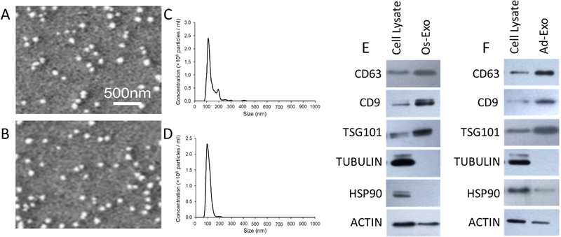
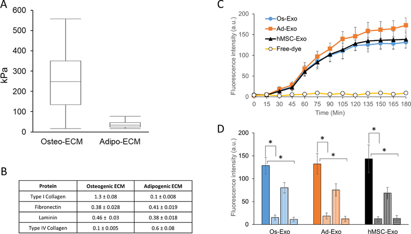
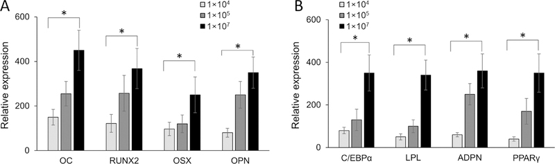
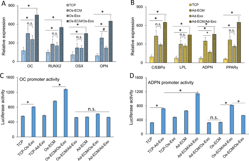
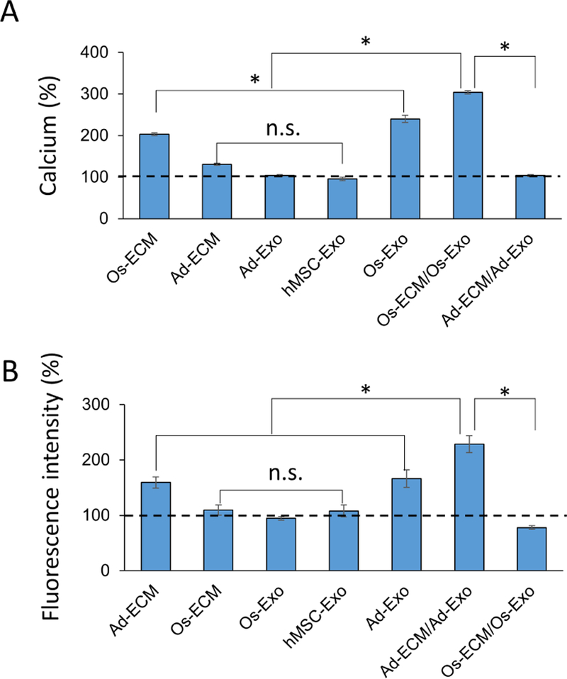
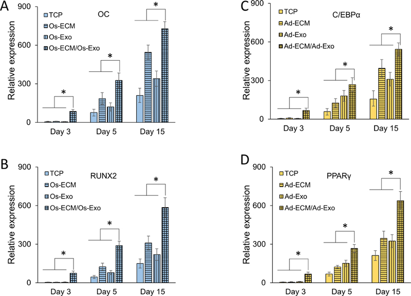
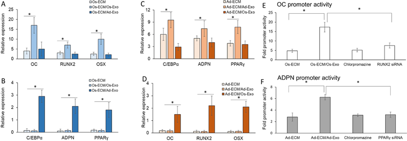
Lineage specification is an essential process in stem cell fate, tissue homeostasis and development. Microenvironmental cues provide direct and selective extrinsic signals to regulate lineage specification of stem cells. Microenvironmental milieu consists of two essential components, one being extracellular matrix (ECM) as the substratum, while the other being cell secreted exosomes and growth factors. ECM of differentiated cells modulates phenotypic expression of stem cells, while their exosomes contain phenotype specific instructive factors (miRNA, RNA and proteins) that control stem cell differentiation. This study demonstrates that osteoblasts-derived (Os-Exo) and adipocytes-derived (Ad-Exo) exosomes contain instructive factors that regulate the lineage specification of human mesenchymal stem cells (hMSCs). Analyses of exosomes revealed the presence of transcription factors in the form of RNA and protein for osteoblasts (RUNX2 and OSX) and adipocytes (C/EBPα and PPARγ). In addition, several miRNAs reported to have osteogenic and adipogenic differentiation potentials are also identified in these exosomes. Kinetic and differentiation analyses indicate that both osteoblast and adipocyte exosomes augment ECM-mediated differentiation of hMSCs into the respective lineage. The combination of osteoblast/adipocyte ECM and exosomes turned-on the lineage specific gene expressions at earlier time points of differentiation compared to the respective ECM or exosomes administered individually. Interestingly, the hMSCs differentiated on osteoblast ECM with adipogenic exosomes showed expression of adipogenic lineage genes, while hMSCs differentiated on adipocyte ECM with osteoblast exosomes showed osteogenic lineage genes. Based on these observations, we conclude that exosomes might override the ECM mediated instructive signals during lineage specification of hMSC.

Keywords: Adipogenesis; Exosomes; Extracellular matrix; Lineage determination; Microenvironment; Osteogenesis; Stem cell fate.


Figures

Figure 1:
Figure 1:. Characterization of exosomes.
Physical parameters of the purified exosomes were analyzed by SEM and NanoSight. Three separate experimental runs were performed for osteoblast and adipocyte derived exosomes. Representative SEM images are presented for osteoblast (A) and adipocyte (B) exosomes. Average size distribution profile of osteoblast (C) and adipocyte (D) exosomes were obtained from the three runs. Cell lysate and exosomes of osteoblast (E) and adipocyte (F) were scrutinized by western blot with specific antibodies.
Figure 2:
Figure 2:. Characterization of cargo of exosomes.
Osteogenic influencing miRNAs (A) and RNAs (B) abundance, and protein (RUNX2, OSX and actin) expression (C) in the purified osteoblast exosomes. Adipogenic influencing miRNAs (D) and RNA (E) abundance, and protein (PPARγ, C/EBPα and actin) expression (F) in the purified adipocyte exosomes. The miRNA and RNA expressions presented represent normalized fluorescent signals (∆Rn) plotted against the number of amplification cycles (A, B, D and E). RUNX2, OSX, PPARγ, C/EBPα and actin expressions were detected by western blot analyses using specific antibodies in respective cell lysates and purified exosomes (Os-Exo/Ad-Exo) (C and F). RUNX2, OC, OSX, PPARγ, C/EBPα, LPL and HSP90 expressions were quantitated by RT-qPCR using gene specific primers. The RNA copy numbers of osteoblasts (G) and adipocytes (H) were estimated using RT-qPCR with known concentrations of respective plasmid DNA. RNAs were isolated from undifferentiated (UD) and differentiated (Diff) at day-7 and day-15 of culture. The copy numbers are presented as mean ± S.D. The RNAs isolated from osteoblast, adipocytes and their respective exosomes were in vitro translated to proteins using cell-free translation system in the presence and absence of RNase. The presence of RUNX2, OSX, PPARγ and C/EBPα were identified by western blot analysis with specific antibodies (I).
Figure 3:
Figure 3:. Characterization of extracellular matrix and exosome internalization.
Normal human osteoblasts (NHO) and pre-adipocytes were differentiated to either osteoblasts or adipocytes on glass coverslips, as described in methods. The lysed cells were aspirated and the stiffness of the selected area (50 µm x 50 µm) deposited extracellular matrix (ECM) was examined under atomic force microscope (AFM). Young’s modulus (A) presented as mean ± S.D. for osteoblast and adipocyte ECM were calculated from 10 randomly selected regions using Hertz model. The extracted ECM was quantitated for specific proteins by ELISA. Type I collagen, fibronectin, laminin and Type IV collagen were measured for three independent extractions and presented as mean ± S.D. (B). Adhered human mesenchymal stem cells (hMSCs) were incubated with either labeled exosomes or free-dye for 24 h, as described in Materials and Methods section. Following incubation, the cells were washed with PBS, fixed with paraformaldehyde and fluorescence intensity was measured in a plate reader. Exosome uptake is presented as mean ± S.D. of fluorescence intensity of osteoblast (Os-Exo), adipocyte (Ad-Exo) and hMSC (hMSC-Exo). The hMSCs were supplemented with labeled exosomes for 2 h either at 37°C (solid bar) in the presence of 5µM (vertical lines bars) and 10µM (horizontal lines bars) of chlorpromazine. While incubation of hMSCs with exosomes at 4°C (slanted line bars) inhibited the uptake of exosomes. One-way ANOVA was used to evaluate statistical significance between groups (*p ≤ 0.005).
Figure 4:
Figure 4:. Effect of exosome concentrations on osteogenic and adipogenic gene expression in human mesenchymal stem cells.
Human mesenchymal stem cells (hMSC) were differentiated on either osteoblast (A) or adipocyte (B) extracellular matrix (ECM). The differentiation on ECM was further supplemented with different concentrations of respective exosomes. After 15 days of differentiation, the osteogenic [OC, RUNX2, OSX and OPN) and adipogenic (C/EBPα, LPL, ADPN and PPARγ) specific gene expressions were quantitated by RT-qPCR analyses using specific primers. Student t-test was used to evaluate the statistical significance (*p ≤ 0.005).
Figure 5:
Figure 5:. Effect of extracellular matrix and exosomes on gene expression in human mesenchymal stem cell differentiation.
Human mesenchymal stem cells (hMSCs) were differentiated in either tissue culture plate (TCP, open bars) or cell type specific extracellular matrix (ECM, vertical line bars). The hMSCs differentiated in TCP (TCP/Exo, horizontal line bars) and ECM (ECM/Exo, checkered bars) were further differentiated in the presence of either osteoblast exosomes (A) or adipocyte exosomes (B). Relative gene expressions presented represent fold changes with respect to undifferentiated hMSCs. Osteogenic (OC, RUNX2, OSX and OPN) and adipogenic (C/EBPα, LPL, ADPN and PPARγ) genes were quantitated by RT-qPCR using gene specific primers. The OC (C) and ADPN (D) promoter activities were assessed during differentiation of hMSCs into osteoblast and adipocytes at different conditions, respectively. The hMSCs grown on either TCP or ECM were supplemented with exosomes as described in Materials and Methods section. The OC and ADPN promoter constructs were transfected individually into the hMSCs on day-15 of differentiation towards osteogenic and adipogenic lineage, respectively. Following 24 h transfection, luciferase activity was measured in the cell lysates. One-way ANOVA was used to evaluate statistical significance between groups (*p ≤ 0.005).
Figure 6:
Figure 6:. Effect of cell specific exosomes on calcium deposition (osteogenesis) and lipid droplets (adipogenesis) on human mesenchymal stem cells.
The human mesenchymal stem cells (hMSCs) were differentiated on either tissue culture plate (TCP) or extracellular matrix (ECM). During osteogenic differentiation on osteoblast ECM (Os-ECM), the cells were also supplemented and differentiated in the presence of osteoblast exosomes (Os-ECM/Os-Exo). The deposited calcium was quantitated using calcium assay kit, normalized to protein and presented as percentage with levels on TCP as 100% (dotted line) (A). During adipogenic differentiation on adipocyte ECM (Ad-ECM), the cells were also supplemented and differentiated in the presence of adipocyte exosomes (Ad-ECM/Ad-Exo). The fluorescent intensity of the lipid droplets was measured at 505 nm, normalized to protein and presented as percentage with levels on TCP as 100% (dotted line) (B). One-way ANOVA was used to evaluate the statistical significance (*p ≤ 0.005).
Figure 7:
Figure 7:. Effect of cell specific exosomes on the kinetics of human mesenchymal stem cell differentiation.
Human mesenchymal stem cell (hMSCs) were differentiated on either tissue culture plate (TCP) (solid bars) or extracellular matrix (ECM; vertical line bars). Exosomes were supplemented during differentiation (TCP/Exo; horizontal line bars) on ECM (ECM/Exo; checkered bars). Following 3, 5 and 15 days of induction, the osteogenic [OC (A) and RUNX2 (B)] and adipogenic [C/EBPα (C) and PPARγ (D)] gene expressions were quantitated by RT-qPCR analyses using specific primers. One-way ANOVA was used to evaluate the statistical significance (*p ≤ 0.005).
Figure 8:
Figure 8:. Effect of cell specific exosomes on extracellular matrix directed human mesenchymal stem cell lineage.
Human mesenchymal stem cells (hMSCs) were differentiated on either osteoblast (Os-ECM) or adipocyte (Ad-ECM) extracellular matrix (ECM). During osteogenic differentiation on Os-ECM the cells were supplemented with either osteoblast exosomes [Os-ECM/Os-Exo; (A)] or adipocyte exosomes [Os-ECM/Ad-Exo (B)], while during adipogenic differentiation on Ad-ECM the cells were supplemented with either osteoblast exosomes [Ad-ECM/Os-Exo (C)] or adipocyte exosomes [Ad-ECM/Ad-Exo (B)]. After 15 days of differentiation the osteogenic [OC, RUNX2 and OSX) and adipogenic (C/EBPα, ADPN and PPARγ) specific gene expressions were quantitated by RT-qPCR analyses using specific primers. The OC (E) and ADPN (F) promoter activities were assessed during the differentiation of hMSCs to osteoblast or adipocyte on either Os-ECM or Ad-ECM with respective exosomes. The promoter activities were also measured in the presence of 10 µM chlorpromazine supplemented with exosomes. The siRNAs targeting RUNX2 and PPARγ were transfected 8 h prior to supplementation of exosomes. One-way Anova was used to evaluate the statistical significance (*p ≤ 0.005).

Similar articles

Cited by

References

    1. Caplan AI. Mesenchymal stem cells. Journal of orthopaedic research 1991;9(5):641–50. - PubMed
    1. Pittenger MF, Mackay AM, Beck SC, Jaiswal RK, Douglas R, Mosca JD, et al. Multilineage potential of adult human mesenchymal stem cells. science 1999;284(5411):143–7. - PubMed
    1. McBeath R, Pirone DM, Nelson CM, Bhadriraju K, Chen CS. Cell shape, cytoskeletal tension, and RhoA regulate stem cell lineage commitment. Developmental cell 2004;6(4):483–95. - PubMed
    1. Narayanan K, Leck K-J, Gao S, Wan AC. Three-dimensional reconstituted extracellular matrix scaffolds for tissue engineering. Biomaterials 2009;30(26):4309–17. - PubMed
    1. Guilak F, Cohen DM, Estes BT, Gimble JM, Liedtke W, Chen CS. Control of stem cell fate by physical interactions with the extracellular matrix. Cell stem cell 2009;5(1):17–26. - PMC - PubMed

Publication types