

Automatic evaluation of tumor budding in immunohistochemically stained colorectal carcinomas and correlation to clinical outcome
- PMID: 30153844
- PMCID: PMC6114534
- DOI: 10.1186/s13000-018-0739-3
Automatic evaluation of tumor budding in immunohistochemically stained colorectal carcinomas and correlation to clinical outcome
Abstract
Background: Tumor budding, meaning a detachment of tumor cells at the invasion front of colorectal carcinoma (CRC) into single cells or clusters (<=5 tumor cells), has been shown to correlate to an inferior clinical outcome by several independent studies. Therefore, it has been discussed as a complementary prognostic factor to the TNM staging system, and it is already included in national guidelines as an additional prognostic parameter. However, its application by manual evaluation in routine pathology is hampered due to the use of several slightly different assessment systems, a time-consuming manual counting process and a high inter-observer variability. Hence, we established and validated an automatic image processing approach to reliably quantify tumor budding in immunohistochemically (IHC) stained sections of CRC samples.
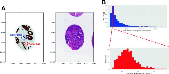
Methods: This approach combines classical segmentation methods (like morphological operations) and machine learning techniques (k-means and hierarchical clustering, convolutional neural networks) to reliably detect tumor buds in colorectal carcinoma samples immunohistochemically stained for pan-cytokeratin. As a possible application, we tested it on whole-slide images as well as on tissue microarrays (TMA) from a clinically well-annotated CRC cohort.
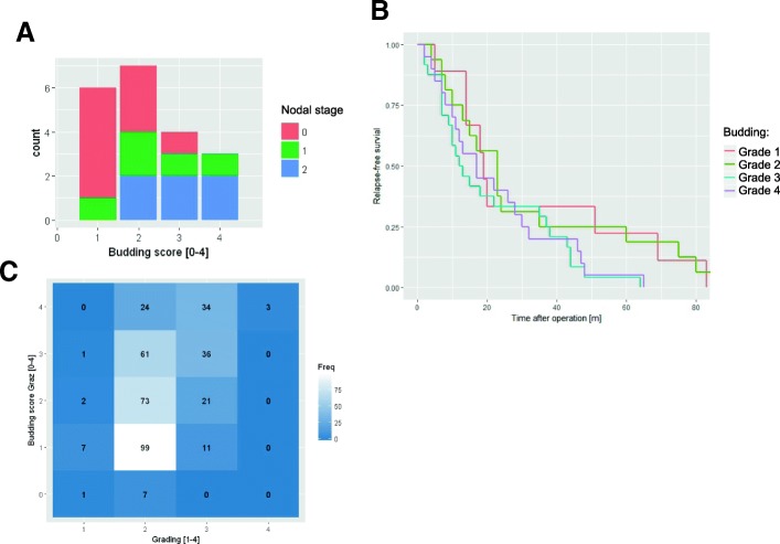
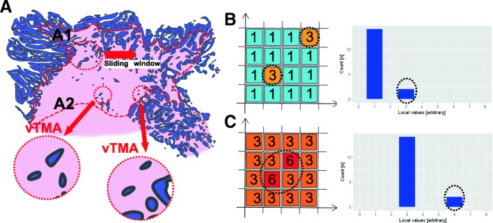
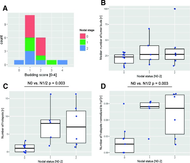
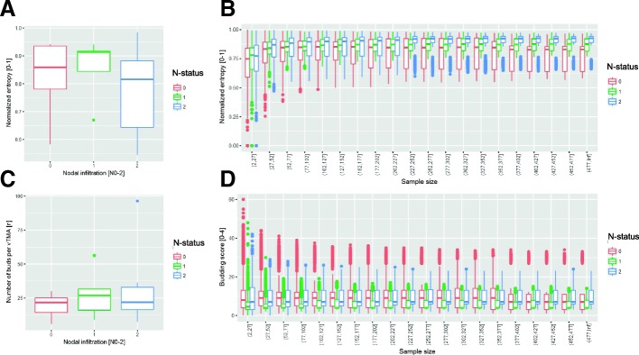
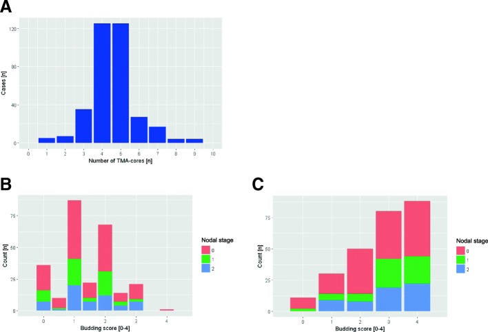
Results: Our automatic tumor budding evaluation tool detected the absolute number of tumor buds per image with a very good correlation to the manually segmented ground truth (R2 value of 0.86). Furthermore the automatic evaluation of whole-slide images from 20 CRC-patients, we found that neither the detected number of tumor buds at the invasion front nor the number in hotspots was associated with the nodal status. However, the number of spatial clusters of tumor buds (budding hotspots) significantly correlated to the nodal status (p-value = 0.003 for N0 vs. N1/N2). TMAs were not feasible for tumor budding evaluation, as the spatial relationship of tumor buds (especially hotspots) was not preserved.
Conclusions: Automatic image processing is a feasible and valid assessment tool for tumor budding in CRC on whole-slide images. Interestingly, only the spatial clustering of the tumor buds in hotspots (and especially the number of hotspots) and not the absolute number of tumor buds showed a clinically relevant correlation with patient outcome in our data.
Keywords: Colorectal carcinoma; Convolutional neural network; Digital pathology; Image processing; Tumor budding.
Conflict of interest statement
Ethics approval and consent to participate
All experiments were in accordance with the local ethics committee (Ethics Committee, Medical University Graz, Austria; decision 18–199 ex 06/07).All patients agreed that their stored material was enclosed and that their clinical data were anonymously used for statistical analysis (as already done in former published studies on this material [9, 10]).
Consent for publication
Not applicable.
Competing interests
The authors declare that they have no competing interests.
Publisher’s Note
Springer Nature remains neutral with regard to jurisdictional claims in published maps and institutional affiliations.
Figures
Similar articles
-
Digital image analysis of pan-cytokeratin stained tumor slides for evaluation of tumor budding in pT1/pT2 colorectal cancer: Results of a feasibility study.Pathol Res Pract. 2018 Sep;214(9):1273-1281. doi: 10.1016/j.prp.2018.07.002. Epub 2018 Jul 10. Pathol Res Pract. 2018. PMID: 30017334
-
Immunohistochemical evaluation of tumor budding for stratifying T1 colorectal cancer: optimal cut-off value and a novel computer-assisted semiautomatic method.Mod Pathol. 2019 May;32(5):675-683. doi: 10.1038/s41379-018-0189-1. Epub 2018 Dec 14. Mod Pathol. 2019. PMID: 30552417
-
Comprehensive assessment of tumour budding by cytokeratin staining in colorectal cancer.Histopathology. 2017 Jun;70(7):1044-1051. doi: 10.1111/his.13164. Epub 2017 Mar 2. Histopathology. 2017. PMID: 28061021
-
Tumor budding in colorectal cancer--ready for diagnostic practice?Hum Pathol. 2016 Jan;47(1):4-19. doi: 10.1016/j.humpath.2015.08.007. Epub 2015 Sep 3. Hum Pathol. 2016. PMID: 26476568 Review.
-
Tumor Budding in Colorectal Carcinoma: Translating a Morphologic Score Into Clinically Meaningful Results.Arch Pathol Lab Med. 2018 Aug;142(8):952-957. doi: 10.5858/arpa.2018-0082-RA. Arch Pathol Lab Med. 2018. PMID: 30040461 Review.
Cited by
-
Application of artificial intelligence to the diagnosis and therapy of colorectal cancer.Am J Cancer Res. 2020 Nov 1;10(11):3575-3598. eCollection 2020. Am J Cancer Res. 2020. PMID: 33294256 Free PMC article. Review.
-
Predictive value of tumor budding in head and neck squamous cell carcinoma: an update.Virchows Arch. 2023 Oct;483(4):441-449. doi: 10.1007/s00428-023-03630-6. Epub 2023 Aug 29. Virchows Arch. 2023. PMID: 37642731 Review.
-
Semi-Supervised Learning to Automate Tumor Bud Detection in Cytokeratin-Stained Whole-Slide Images of Colorectal Cancer.Cancers (Basel). 2023 Mar 30;15(7):2079. doi: 10.3390/cancers15072079. Cancers (Basel). 2023. PMID: 37046742 Free PMC article.
-
Breast Cancer Classification Based on Tumor Budding and Stem Cell-Related Signatures Facilitate Prognosis Evaluation.Front Oncol. 2022 Jan 10;11:818869. doi: 10.3389/fonc.2021.818869. eCollection 2021. Front Oncol. 2022. PMID: 35083162 Free PMC article.
-
Deep Learning on Histopathological Images for Colorectal Cancer Diagnosis: A Systematic Review.Diagnostics (Basel). 2022 Mar 29;12(4):837. doi: 10.3390/diagnostics12040837. Diagnostics (Basel). 2022. PMID: 35453885 Free PMC article. Review.
References
-
- WHO . Classification of Tumors of the Digestive System. 4 2010.
-
- Wittekind C. TNM: Klassifikation maligner Tumoren: Wiley; 2017.
-
- Gospodarowicz MK, Brierley JD, Wittekind C. TNM classification of malignant tumors: Wiley; 2017.
Publication types
MeSH terms
Substances
Grants and funding
LinkOut - more resources
Full Text Sources
Other Literature Sources
Medical
Research Materials
