. 2018 Oct 12;293(41):15855-15866.
doi: 10.1074/jbc.RA118.004806. Epub 2018 Aug 28.

Structural analysis reveals pathomechanisms associated with pseudoxanthoma elasticum-causing mutations in the ABCC6 transporter

Affiliations

Structural analysis reveals pathomechanisms associated with pseudoxanthoma elasticum-causing mutations in the ABCC6 transporter

Yanchao Ran et al. J Biol Chem. .

Abstract

Mutations in ABC subfamily C member 6 (ABCC6) transporter are associated with pseudoxanthoma elasticum (PXE), a disease resulting in ectopic mineralization and affecting multiple tissues. A growing number of mutations have been identified in individuals with PXE. For most of these variants, no mechanistic information is available regarding their role in normal and pathophysiologies. To assess how PXE-associated mutations alter ABCC6 biosynthesis and structure, we biophysically and biochemically evaluated the N-terminal nucleotide-binding domain. A high-resolution X-ray structure of nucleotide-binding domain 1 (NBD1) of human ABCC6 was obtained at 2.3 Å that provided a template on which to evaluate PXE-causing mutations. Biochemical analysis of mutations in this domain indicated that multiple PXE-causing mutations altered its structural properties. Analyses of the full-length protein revealed a strong correlation between the alterations in NBD properties and the processing and expression of ABCC6. These results suggest that a significant fraction of PXE-associated mutations located in NBD1 causes changes in its structural properties and that these mutation-induced alterations directly affect the maturation of the full-length ABCC6 protein.

Keywords: ABC transporter; ABCC6; X-ray crystallography; connective tissue; connective tissue disorder; elastic fiber; glycoprotein biosynthesis; membrane protein; protein folding; protein misfolding; protein stability; protein structure; pseudoxanthoma elasticum.


Conflict of interest statement

The authors declare that they have no conflicts of interest with the contents of this article

Figures
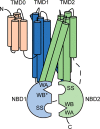
Figure 1.
Figure 1.
ABCC6 structure. A cartoon representation of the ABCC6 structure is shown and colored by domain. The canonical Walker A (WA), Walker B (WB), and signature (SS) sequences are indicated. The degenerate Walker B site in NBD1 is indicated with WB*.
Figure 2.
Figure 2.
ABCC subfamily NBD1 alignment. A multiple sequence alignment for the human NBD1 ABCC domains is shown. Conserved sequence elements associated with ATP binding and hydrolysis are indicated below the alignment. The secondary structure assignments from the ABCC6 NBD1 structure are shown above the alignment with arrows indicating strands and curling loops indicating helices. The secondary structures symbols are colored by subdomain assignment (β-sheet, yellow; α/β-core, purple; α-helical, green). Secondary structure numbering was adapted from the HisP NBD structure (PDB code 1B0U) (44). The figure was prepared with ESPRIPT and BoxShade (45).
Figure 3.
Figure 3.
NBD1 structure. The structure and topology of ABCC6 NBD1 is shown. A, a cartoon representation of ABCC6 NBD1 is shown in two orientations rotated by 60°. The subdomains are colored as in Fig. 2 (β-sheet, yellow; α/β-core, purple; α-helical, green). The structurally diverse region, part of the α-helical subdomain, is shown (tan). B, a cartoon representation of ABCC6 NBD1 showing the location of the canonical ATP-binding sequences is shown in two orientations, as in A. The labels of the conserved elements are colored corresponding to the features in the structure. C, a tube diagram of NBD1 is shown colored and scaled by B-factor. D, a topology map of ABCC6 NBD1 is shown colored as in A.
Figure 4.
Figure 4.
NBD1 subdomain orientation, ATP binding, and catalytic site in ABCC structures. The structures of other ABCC NBD1 proteins were superposed with that of ABCC6 NBD1 using the conserved, α/β-core Walker A and B sequences. A, superposition of ABCC1/MRP1 structures with ABCC6 NBD1 is shown as a ribbon with ABCC6 (green), nucleotide-free ABCC1/MRP1 (4C3Z, yellow), and ATP-bound ABCC1/MRP1 (2CBZ, cyan) solved as isolated domains. ABCC1/MRP1 NBD1 from the full-length cryo-EM structure is also shown in the ATP-bound state (PDB code 6BHU, purple). B, superposition of ABCC6 NBD1 with NBD1 (green) from the full-length cryo-EM structure of ABCC8/SUR1 (PDB code 6BAA, brown) is shown, aligned as in A. C, key binding and catalytic residues in ABCC6 NBD1 are shown as side-chain sticks. The experimentally observed sulfate ion is shown as partially transparent spheres. The modeled nucleotide and Mg2+ ion are shown as sticks and a blue sphere, respectively. D, comparison of the active sites from the ABCC NBD1 structures is shown. The region shown in D is boxed in C for reference. The H-loop histidine adopts multiple conformations across the NBD structures: a, ABCC6 NBD1; b, 6BHU: ABCC1/MRP1, full-length, ATP-bound; c, 4C3Z: ABCC6/MRP1, domain, apo; d, 5UJ9: ABCC1/MRP1, full-length, ATP-bound; e, 2CBZ: ABCC1/MRP1, domain, ATP-bound; f, ABCC8/SUR1, full-length, apo. The Walker A (blue), Walker B (purple), and signature sequences (brown) are colored for reference.
Figure 5.
Figure 5.
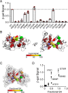
NBD1 expression. NBD expression was assessed using an enzymatic assay in mammalian cells. A, the effects of PXE mutations are shown on NBD1 expression, normalized to the signal from WT. B, the effects of PXE mutations are mapped onto the structure of ABCC6 NBD1 and colored by severity. Two views of the NBD1 are shown rotated by 60°. C, the position of the PXE mutations are shown and colored by fractional surface area. The partially transparent molecular surface is shown as a reference. D, the correlation between fractional surface area and NBD1 expression is shown. Variants showing intermediate or greater expression are labeled for reference. The data shown are means ± S.D. from n > 4 experiments. Individual data points used for the analyses are shown for each mutant.
Figure 6.
Figure 6.
ABCC6 expression. PXE-associated mutations in NBD1 were assessed for their impact on the folding and maturation of full-length ABCC6 in cell culture. A, a representative Western blot is shown. Molecular mass markers are indicated on the left (in kDa), and the positions of the core (band B) and complexly glycosylated (band C) protein species are shown on the right. PARP1 is shown as a loading control. A dark exposure was chosen to demonstrate expression of all ABCC6 clones. B, densitometric analysis of Western blotting of ABCC6 expression is shown. The data shown are means ± S.E. from n > 4 experiments. Individual data points used for the analyses are shown for each mutant.
Figure 7.
Figure 7.
NBD1 expression and ABCC6 trafficking. The correlation between NBD expression and full-length trafficking was assessed from data presented in Figs. 5 and 6. NBD1 expression from the β-gal assay is plotted against the steady-state expression of full-length ABCC6. Residues showing intermediate or greater levels of expression are labeled. Residues located in conserved ATP-binding and hydrolysis sequences are shown in green (Q698P, E699D, and G755R). Unity is shown as a dashed line for reference.

References

    1. Goodman R. M., Smith E. W., Paton D., Bergman R. A., Siegel C. L., Ottesen O. E., Shelley W. M., Pusch A. L., and McKusick V. A. (1963) Pseudoxanthoma elasticum: a clinical and histopathological study. Medicine 42, 297–334 10.1097/00005792-196309000-00001 - DOI - PubMed
    1. Bergen A. A., Plomp A. S., Schuurman E. J., Terry S., Breuning M., Dauwerse H., Swart J., Kool M., van Soest S., Baas F., ten Brink J. B., and de Jong P. T. (2000) Mutations in ABCC6 cause pseudoxanthoma elasticum. Nat. Genet. 25, 228–231 10.1038/76109 - DOI - PubMed
    1. Le Saux O., Urban Z., Tschuch C., Csiszar K., Bacchelli B., Quaglino D., Pasquali-Ronchetti I., Pope F. M., Richards A., Terry S., Bercovitch L., de Paepe A., and Boyd C. D. (2000) Mutations in a gene encoding an ABC transporter cause pseudoxanthoma elasticum. Nat. Genet. 25, 223–227 10.1038/76102 - DOI - PubMed
    1. Jansen R. S., Küçükosmanoglu A., de Haas M., Sapthu S., Otero J. A., Hegman I. E., Bergen A. A., Gorgels T. G., Borst P., and van de Wetering K. (2013) ABCC6 prevents ectopic mineralization seen in pseudoxanthoma elasticum by inducing cellular nucleotide release. Proc. Natl. Acad. Sci. U.S.A. 110, 20206–20211 10.1073/pnas.1319582110 - DOI - PMC - PubMed
    1. Iliás A., Urbán Z., Seidl T. L., Le Saux O., Sinkó E., Boyd C. D., Sarkadi B., and Váradi A. (2002) Loss of ATP-dependent transport activity in pseudoxanthoma elasticum-associated mutants of human ABCC6 (MRP6). J. Biol. Chem. 277, 16860–16867 10.1074/jbc.M110918200 - DOI - PubMed

Publication types

MeSH terms

Substances

LinkOut - more resources