Review
. 2018 Aug 28;7(9):121.
doi: 10.3390/cells7090121.

Centrosomal and Non-Centrosomal Microtubule-Organizing Centers (MTOCs) in Drosophila melanogaster

Affiliations
Review

Centrosomal and Non-Centrosomal Microtubule-Organizing Centers (MTOCs) in Drosophila melanogaster

Marisa M L Tillery et al. Cells. .

Abstract

The centrosome is the best-understood microtubule-organizing center (MTOC) and is essential in particular cell types and at specific stages during Drosophila development. The centrosome is not required zygotically for mitosis or to achieve full animal development. Nevertheless, centrosomes are essential maternally during cleavage cycles in the early embryo, for male meiotic divisions, for efficient division of epithelial cells in the imaginal wing disc, and for cilium/flagellum assembly in sensory neurons and spermatozoa. Importantly, asymmetric and polarized division of stem cells is regulated by centrosomes and by the asymmetric regulation of their microtubule (MT) assembly activity. More recently, the components and functions of a variety of non-centrosomal microtubule-organizing centers (ncMTOCs) have begun to be elucidated. Throughout Drosophila development, a wide variety of unique ncMTOCs form in epithelial and non-epithelial cell types at an assortment of subcellular locations. Some of these cell types also utilize the centrosomal MTOC, while others rely exclusively on ncMTOCs. The impressive variety of ncMTOCs being discovered provides novel insight into the diverse functions of MTOCs in cells and tissues. This review highlights our current knowledge of the composition, assembly, and functional roles of centrosomal and non-centrosomal MTOCs in Drosophila.

Keywords: Drosophila; centriole; centrosome; microtubule; microtubule-organizing center (MTOC); ninein; non-centrosomal MTOC; patronin; γ-tubulin.


Conflict of interest statement

The authors declare no conflict of interest.

Figures

Figure 1
Figure 1
Structure of the Drosophila centrosome. The organization of several centriolar and pericentriolar material (PCM) proteins in the interphase centrosome. The mother centriole organizes PCM, shown as three layers, and maintains a tight association (engagement) with the daughter centriole. The figure is based on models presented in [16,17,18].
Figure 2
Figure 2
Centriole duplication cycle. Centriole pairs disengage in late mitosis, licensing mother and daughter centrioles to duplicate. Procentriole (grey) genesis begins with cartwheel (orange) assembly at the base of the old and new mother centrioles (blue) as cells enter S phase. Procentrioles elongate during S and G2 phases. During mitosis, DNA (purple) is segregated to each daughter cell through the activity of the centrosomes. PCM is recruited and the procentriole is converted to a mature mother capable of duplicating once the cell passes through mitosis. In late mitosis, the centriole pair disengages and the old and new mothers are licensed to duplicate.
Figure 3
Figure 3
A non-centrosomal microtubule organizing center (ncMTOC) assembles during meiosis II. Meiosis I spindle assembly is anastral, but in late meiosis I a central aster forms, comprised of centrosomal PCM proteins, that separates the two meiosis II spindles. The blue structures represent the central aster ncMTOC; MTs are green; DNA is purple. Figure based on [152].
Figure 4
Figure 4
Centrosomes are essential to organize Rappaport furrows during embryonic cleavage cycles. The dynamics of centrosomes and furrow formation during cleavage cycles. Figure based on [174].
Figure 5
Figure 5
Spermatogenesis. Spermatogenic germline stem cells reside at a niche called the hub where divisions are polarized and asymmetric, producing spermatogonia. Spermatogonia divide four times to produce a cyst of 16 spermatocytes. Spermatocytes assemble short cilia from each of their four centrioles during G2 phase and retain them as they undergo two meiotic divisions to produce sixty-four haploid spermatids. Spermatids assemble an axoneme in their cytoplasm and the mitochondria fuse into two large mitochondrial derivatives upon which ncMTOCs assemble. Nuclei are grey; MTs are green; mitochondria are brown; MTOCs (centrosomes and ncMTOCs on the mitochondrial derivative) are blue.
Figure 6
Figure 6
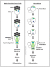
Male germline stem cells (GSCs) and neuroblasts exhibit centrosome asymmetry. Male germline stem cells retain the older mother centriole, which anchors at the apical membrane. Neuroblasts, on the other hand, anchor the daughter centrosome at the apical cell membrane. The neuroblast daughter centrosome maintains active MTOC activity during interphase, while the mother loses it until mitosis. Hub cells in the niche are dark grey; procentrioles are grey; centrioles with MTOC activity are blue: the daughter centrosome is light blue, the mother centrosome is dark blue.
Figure 7
Figure 7
Organization of the ncMTOC in ovarian follicle cells. Shot, not depicted in the illustration, is associated with the apical spectrin cytoskeleton as is the ncMTOC.
Figure 8
Figure 8
Salivary gland placode cell ncMTOC. In this cell type, the MTOC changes dynamically during embryonic stage 11 from a centrosomal MTOC to an apical membrane ncMTOC required for morphogenesis.
Figure 9
Figure 9
An apical membrane ncMTOC assembles in tracheal epithelia. The MTOC is blue and transitions from the centrosome to an apical ncMTOC; MTs are green; the nucleus is grey.
Figure 10
Figure 10
Wing epithelial cell ncMTOC. During larval and early pupal stages (AC), wing epithelial cells employ an apical junction to assemble a dynamic ncMTOC along the proximal–distal axis that controls planar cell polarity. By 30 h into pupal development (D), the MT array loses its ordered proximal–distal organization. At the final stages of wing morphogenesis (E), a ncMTOC forms at the apical surface of trichome-bearing cells and organizes MT arrays along the apical–basal axis. The ncMTOC is blue; MTs are green; nuclei and centrosomes are grey.
Figure 11
Figure 11
MTs are organized in photoreceptor cells near the apical membrane where the rhabdomere forms. This set of MTs appear to be organized from an undefined ncMTOC.
Figure 12
Figure 12
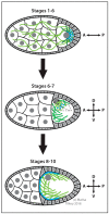
Dynamic changes in MTOCs during oocyte development. In stages 1–6, a cluster of centrioles at the oocyte posterior organizes an MTOC together with a ncMTOC assembled on the posterior hemisphere of the oocyte nucleus. At stage 7, the oocyte migrates to the anterior-dorsal corner of the oocyte and then a new ncMTOC assembles on the anterior cortex. Nuclei are darker grey; MTs are green; centrioles (dots) and other MTOCs are blue.
Figure 13
Figure 13
Perinuclear ncMTOC in muscle cells. The regular spacing of nuclei in multinucleate muscle cells requires the perinuclear ncMTOC.
Figure 14
Figure 14
Neurons assemble ncMTOCs within dendrite branches. Neuronal ncMTOCs regulate dendrite branching. The nucleus is dark grey; MTs are green; ncMTOCs are blue.
Figure 15
Figure 15
A splice variant of Cnn assembles ncMTOCs on the surface of spermatid mitochondria. During spermatid differentiation and elongation, CnnT localizes to mitochondrial derivatives, recruits γ-TuRCs, and converts mitochondria to ncMTOCs.

References

    1. Cooper G.M., Hausman R.E. The Cell: A Molecular Approach. 6th ed. Sinauer Associates, Inc.; Sunderland, MA, USA: 2013.
    1. Sanchez A.D., Feldman J.L. Microtubule-organizing centers: From the centrosome to non-centrosomal sites. Curr. Opin. Cell Biol. 2017;44:93–101. doi: 10.1016/j.ceb.2016.09.003. - DOI - PMC - PubMed
    1. Wu J., Akhmanova A. Microtubule-organizing centers. Annu. Rev. Cell Dev. Biol. 2017;33:51–75. doi: 10.1146/annurev-cellbio-100616-060615. - DOI - PubMed
    1. Baas P.W., Lin S. Hooks and comets: The story of microtubule polarity orientation in the neuron. Dev. Neurobiol. 2011;71:403–418. doi: 10.1002/dneu.20818. - DOI - PMC - PubMed
    1. Akhmanova A., Steinmetz M.O. Control of microtubule organization and dynamics: Two ends in the limelight. Nat. Rev. Mol. Cell Biol. 2015;16:711–726. doi: 10.1038/nrm4084. - DOI - PubMed

LinkOut - more resources