. 2018 Aug 28;8(1):12945.
doi: 10.1038/s41598-018-30964-y.

Corneal myofibroblasts inhibit regenerating nerves during wound healing

Affiliations

Corneal myofibroblasts inhibit regenerating nerves during wound healing

Kye-Im Jeon et al. Sci Rep. .

Abstract

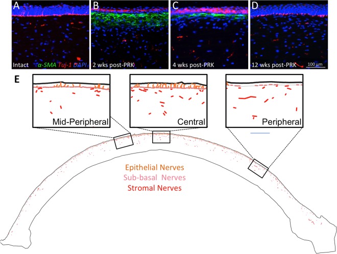
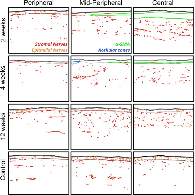
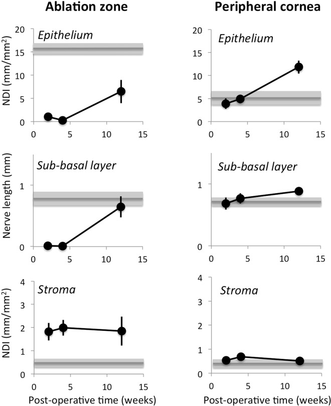
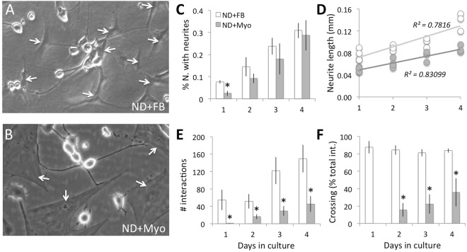
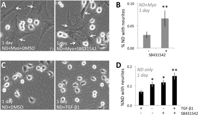
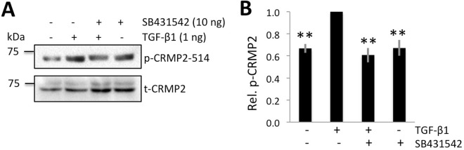
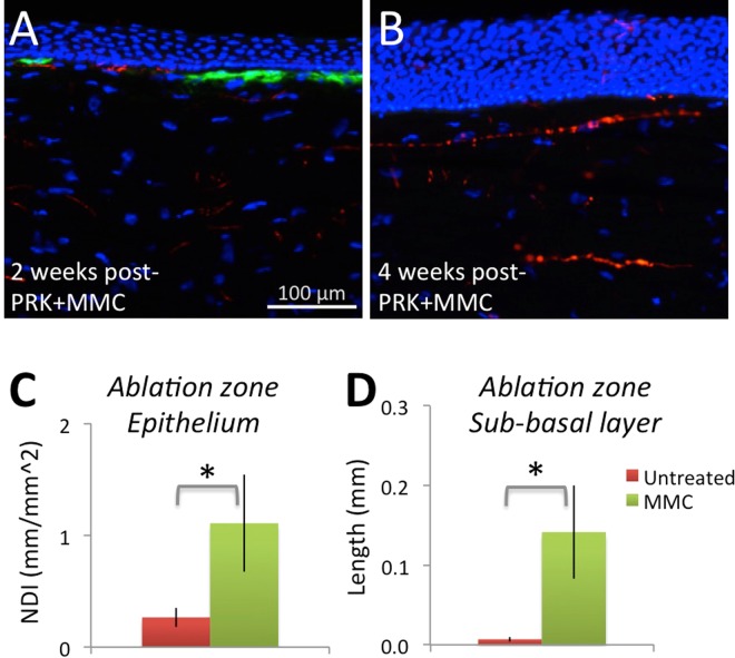
Abnormal nerve regeneration often follows corneal injury, predisposing patients to pain, dry eye and vision loss. Yet, we lack a mechanistic understanding of this process. A key event in corneal wounds is the differentiation of keratocytes into fibroblasts and scar-forming myofibroblasts. Here, we show for the first time that regenerating nerves avoid corneal regions populated by myofibroblasts in vivo. Recreating this interaction in vitro, we find neurite outgrowth delayed when myofibroblasts but not fibroblasts, are co-cultured with sensory neurons. After neurites elongated sufficiently, contact inhibition was observed with myofibroblasts, but not fibroblasts. Reduced neurite outgrowth in vitro appeared mediated by transforming growth factor beta 1 (TGF-β1) secreted by myofibroblasts, which increased phosphorylation of collapsin response mediating protein 2 (CRMP2) in neurons. The significance of this mechanism was further tested by applying Mitomycin C after photorefractive keratectomy to decrease myofibroblast differentiation. This generated earlier repopulation of the ablation zone by intra-epithelial and sub-basal nerves. Our findings suggest that attaining proper, rapid corneal nerve regeneration after injury may require blocking myofibroblast differentiation and/or TGF-β during wound healing. They also highlight hitherto undefined myofibroblast-neuron signaling processes capable of restricting neurite outgrowth in the cornea and other tissues where scars and nerves co-exist.


Conflict of interest statement

The authors declare no competing interests.

Figures

Figure 1
Figure 1
Immunohistochemical staining and analysis of feline corneas. (A) Photograph of normal, unoperated central cat corneal cross-section reacted for Tuj-1 (red fluorescence), α-SMA (green fluorescence) and counter-stained with DAPI (blue fluorescence). Note thick cords of Tuj-1 positive corneal nerves in the anterior stroma, the almost continuous sub-basal nerves right under the epithelium and the thin nerve endings visible between epithelial cells. (B) Photograph of the central cornea of a cat 2 weeks after PRK stained identically as in A. Note the distinct zone of positive α-SMA staining which lacks Tuj-1 positive nerves, the thinner but more densely distributed stromal nerves under that zone, and the Tuj-1 positive epithelium above that zone, which is devoid of intra-epithelial nerves. Note also the lack of sub-basal nerves. (C) Photograph of the central cornea of a cat 4 weeks after PRK, stained identically as in A and B. Note the thinner α-SMA positive zone, which remains devoid of nerves, in spite of their presence below it. The epithelium has increased in thickness, but also remains devoid of nerves. (D) Photograph of the central cornea in a cat 12 weeks after PRK showing a complete lack of α-SMA positive staining. Thicker trunks of Tuj-1 positive nerves are re-appearing in the stroma, as are intra-epithelial and sub-basal nerves. Scale bar = 100 µm for A-D. High-resolution monochrome views of Tuj-1 staining in (A–D) are presented in Fig. S1. (E) Tracing of an entire cat corneal cross section (unoperated cat) performed in Neurolucida, and illustrating with differential color-coding, the different compartments in which corneal nerve densities were analyzed. Insets show higher power views of the central, mid-peripheral and peripheral regions of the cornea.
Figure 2
Figure 2
Sample tracings of immuno-stained corneal sections at different times post-PRK. The 3 columns show illustrative tracings from the peripheral, mid-peripheral and central corneas of 4 different cat eyes: first row, 2 weeks post-PRK; second row, 4 weeks post-PRK, third row, 12 weeks post-PRK and fourth row, unoperated control. Red and orange: Tuj-1 positive corneal nerves in stroma and epithelial layers, respectively. Green: regions positive for α-SMA. Blue: acellular zones. In all cases, the epithelium is shown at the top of each image.
Figure 3
Figure 3
Quantitative analysis of nerve distributions in central and peripheral cat corneas post-PRK. Grey lines and shaded zones indicate mean ± standard error of the mean (SEM) of values obtained in normal, unoperated control corneas (n = 5). NDI: nerve density index. All data points are means ± SEM. See text for statistics.
Figure 4
Figure 4
Differential effect on neurite growth of co-culturing ND7/23 cells with fibroblasts (FB) or myofibroblasts. (A) Phase contrast photograph of plated ND7/23 cells (highly refractile, round cell bodies) and FB (examples arrowed) after 4 days in culture. Note the long, thin neurites expressed by ND cells, which extend liberally across FB cell bodies and processes. (B) Phase contrast photograph of plated ND7/23 cells (highly refractile cell bodies) and Myo (examples arrowed) after 4 days in culture. Note the long, thin neurites expressed by ND cells, which exhibit distinct end-stopping when they contact Myos. (C) Plot of the percentage of plated ND7/23 cells with neurites >40 µm long at different times in either FB or Myo co-culture. While neuritogenesis appears slower in ND + Myo than ND + FB co-cultures at day 1, this difference decreases and disappears on subsequent days. (D) However, total neurite length/cell remained significantly greater in ND + FB co-cultures than in ND + Myo co-cultures at all time-points examined. (E) Plot of the total number of interactions (contacts) between ND cells and either FB or Myos. Note the persistently lower number of interactions between ND7/23 cells and Myos at all time-points, in spite of the fact that the same number of ND cells and Myos were plated initially. (F) Plot of the number of successful cell body crossings as a proportion of the total number of interactions between ND7/23 cells and either FB or Myo on different days in culture. When neurons contact a FB, they cross over its cell body and continue their extension >80% of the time. When ND cells contact a Myo, they tended to stop their advance; no crossings happened during Day 1 of culture, although they increased up to ~40% of the time by Day 4 in culture. All values are means ± SD from 3 separate experiments. *P < 0.05. See Fig. S3A for sample Western blot made from 3 days old co-cultures in this experiment, confirming the presence of Tuj-1 positive cells in both co-culture types, the strong presence of α-SMA positive myofibroblasts in the ND + Myo co-cultures, and their total absence in ND + FB co-cultures.
Figure 5
Figure 5
Critical role of TGF-β1 and its receptor in mediating the inhibitory effect of myofibroblasts (Myos) on neurite outgrowth in 1 day old cultures. (A) Phase contrast photograph of plated ND7/23 cells (highly refractile, round cell bodies) and co-cultured Myos (examples arrowed) in the presence of DMSO or the TGF-β receptor inhibitor SB431542. Note the rare neurites expressed by ND7/23 cells after 1 day in co-culture with Myos and DMSO, in contrast with the numerous neurites expressed by ND cells, in the same co-culture, when SB431542 is present. (B) Plot of the percentage of plated ND7/23 cells with neurites >40 µm long in ND + Myo co-cultures in the presence or absence of SB431542. See Fig. S3B for sample Western blot made from co-cultures in this experiment, confirming the presence of similar amounts of Tuj-1 and of α-SMA positive myofibroblasts irrespective of treatment with SB431542. (C) Phase contrast photograph of ND7/23 cells (highly refractile, round cell bodies) in pure cultures in the presence of DMSO or TGF-β1. Note the multiple neurites expressed by ND7/23 cells after 1 day in culture with DMSO, and the marked decrease in neurites when ND cells are treated with TGF-β1. (D) Plot of the percentage of plated ND7/23 cells with neurites >40 µm long when treated with or without TGF-β1. The dependence of this effect on activation of the TGF-β receptor is demonstrated by the ability of the TGF-β receptor inhibitor SB431542 to eliminate the anti-neuritogenic impact of TGF-β1. See Fig. S3C for sample Western blot made from mono-cultures in this experiment, confirming the presence of similar amounts of Tuj-1 irrespective of treatment with DMSO, TGF-β1, SB431542 or TGF-β1 + SB431542. All values in graphs are means ± SD from 3 separate experiments. * P < 0.05, **P < 0.01.
Figure 6
Figure 6
TGF-β1 increases relative p-CRMP2 expression in pure ND7/23 cultures. (A) Representative Western blots showing protein levels for phosphorylated (p-) CRMP2 and total (t-) CRMP2 in cultured ND7/23 cells with or without TGF-β1 stimulation. The samples were run on separate blots. Basal levels of both p- and t-CRMP2 were distinctly above zero. After 1 hour, TGF-β1 increased the expression of p-CRMP2, but not that of t-CRMP2. The TGF-β receptor inhibitor SB431542 blocked the up-regulation of p-CRMP2 observed following TGF-β1 stimulation. (B) Plot of relative expression of p-CRMP2/t-CRMP2 normalized to densitometric values obtained in cells treated with TGF-β1. Data shown are averaged over three experiments. Total CRMP2 levels were were used as loading controls. Data are expressed as means ± SD. **P < 0.01 versus the TGF-β1 only condition. Full, unedited gels analyzed are shown in Supplementary Figure 5 (Fig. S5).
Figure 7
Figure 7
Immunohistochemical staining and analysis of feline corneas treated with PRK + MMC. (A) Photograph of the central cornea of a cat 2 weeks after PRK + MMC reacted for Tuj-1 (red fluorescence), α-SMA (green fluorescence) and counter-stained with DAPI (blue fluorescence). Note the thin zone of positive α-SMA staining, which is adjacent to a zone devoid of α-SMA staining and which contains nerves. Note also the thin, densely distributed stromal nerves, and the fully-regenerated epithelium. (B) Photograph of the central cornea of a cat 4 weeks after PRK + MMC, stained identically as in A. Note the total absence of an α-SMA positive zone, which contrasts with that seen in Fig. 1. The epithelium is markedly hyperplastic and contains some sparse nerves. (C) Plot of mean ± SEM epithelial NDI (nerve density index) at 4 weeks post-PRK in cat eyes treated with MMC or untreated. There are significantly more corneal nerves in the epithelium following MMC treatment than in untreated corneas (*p < 0.05). (D) Plot of mean ± SEM sub-basal nerve length at 4 weeks post-PRK in cat eyes treated with MMC or untreated. There are almost no sub-basal nerves in the ablation zone of cats with PRK only. There are significantly more corneal nerves in the sub-basal layer of the ablation zone 4 weeks after PRK + MMC treatment (*p < 0.05).

References

    1. Rozsa AJ, Beuerman RW. Density and organization of free nerve endings in the corneal epithelium of the rabbit. Pain. 1982;14:105–120. doi: 10.1016/0304-3959(82)90092-6. - DOI - PubMed
    1. Belmonte C, Acosta MC, Gallar J. Neural basis of sensation in intact and injured corneas. Experimental Eye Research. 2004;78:513–525. doi: 10.1016/j.exer.2003.09.023. - DOI - PubMed
    1. Marfurt CF, Cox J, Deek S, Dvorscak L. Anatomy of the human corneal innervation. Experimental Eye Research. 2010;90:478–492. doi: 10.1016/j.exer.2009.12.010. - DOI - PubMed
    1. Müller LJ, Marfurt CF, Kruse F, Tervo TMT. Corneal nerves: structure, contents and function. Experimental Eye Research. 2003;76:521–542. doi: 10.1016/S0014-4835(03)00050-2. - DOI - PubMed
    1. Shaheen BS, Bakir M, Jain S. Corneal nerves in health and disease. Survey of Ophthalmology. 2014;59:263–285. doi: 10.1016/j.survophthal.2013.09.002. - DOI - PMC - PubMed

Publication types

Substances