. 2018 Aug 28;8(1):12991.
doi: 10.1038/s41598-018-31285-w.

Multifunctional Nanotherapeutics for the Treatment of neuroAIDS in Drug Abusers

Affiliations

Multifunctional Nanotherapeutics for the Treatment of neuroAIDS in Drug Abusers

Rahul Dev Jayant et al. Sci Rep. .

Abstract

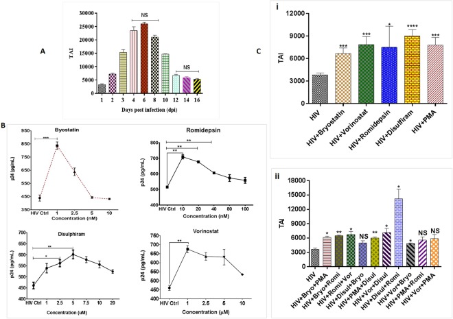
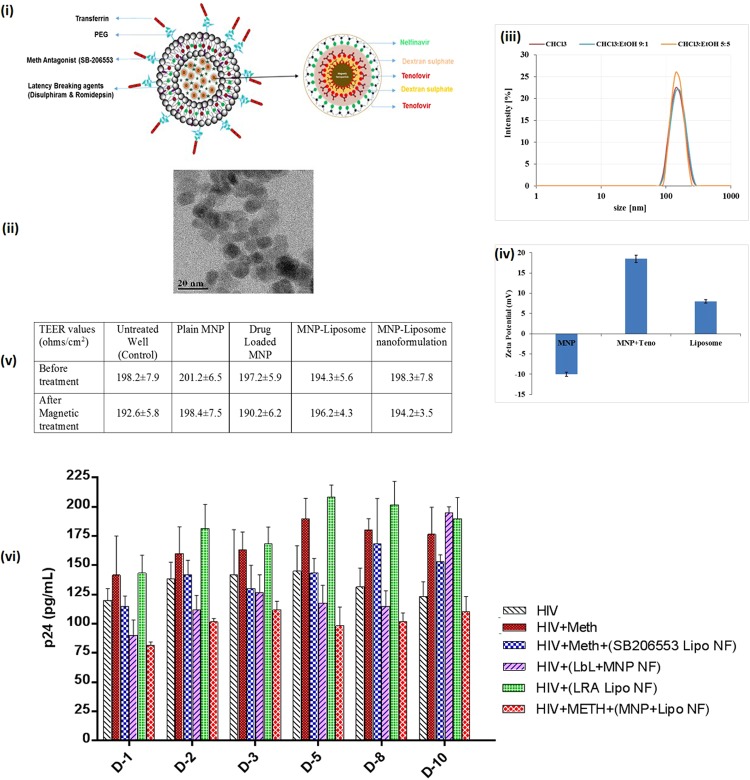
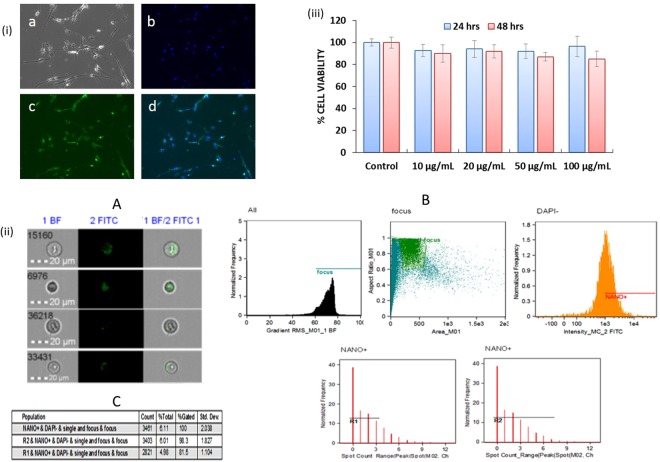
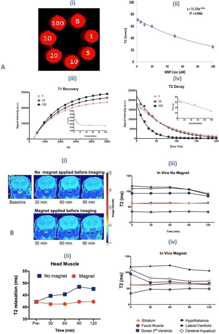
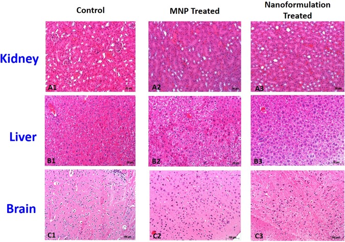
HIV and substance abuse plays an important role in infection and disease progression. Further, the presence of persistent viral CNS reservoirs makes the complete eradication difficult. Thus, neutralizing the drug of abuse effect on HIV-1 infectivity and elimination of latently infected cells is a priority. The development of a multi-component [antiretroviral drugs (ARV), latency reactivating agents (LRA) and drug abuse antagonist (AT)] sustained release nanoformulation targeting the CNS can overcome the issues of HIV-1 cure and will help in improving the drug adherence. The novel magneto-liposomal nanoformulation (NF) was developed to load different types of drugs (LRAs, ARVs, and Meth AT) and evaluated for in-vitro and in-vivo BBB transmigration and antiviral efficacy in primary CNS cells. We established the HIV-1 latency model using human astrocyte cells (HA) and optimized the dose of LRA for latency reversal, Meth AT in in-vitro cell culture system. Further, PEGylated magneto-liposomal NF was developed, characterized for size, shape, drug loading and BBB transport in-vitro. Results showed that drug released in a sustained manner up to 10 days and able to reduce the HIV-1 infectivity up to ~40-50% (>200 pg/mL to <100 pg/mL) continuously using single NF treatment ± Meth treatment in-vitro. The magnetic treatment (0.8 T) was able to transport (15.8% ± 5.5%) NF effectively without inducing any toxic effects due to NF presence in the brain. Thus, our approach and result showed a way to eradicate HIV-1 reservoirs from the CNS and possibility to improve the therapeutic adherence to drugs in drug abusing (Meth) population. In conclusion, the developed NF can provide a better approach for the HIV-1 cure and a foundation for future HIV-1 purging strategies from the CNS using nanotechnology platform.


Conflict of interest statement

The authors declare no competing interests.

Figures

Figure 1
Figure 1
Schematic representation of the development of magneto-liposome NF for drug delivery across the BBB: (A) MNP-LbL assembly of drugs; (B) Magneto-liposome NF preparation; (C) Proposed mechanism of action of NF.
Figure 2
Figure 2
(A) Development of latent HA model; (B) Dose optimization of different LRA agents; (C) Effects of latency reversing agents on provirus quiescence in HA populations (14 dpi), latent HIV cells were treated with optimized dose of (a) Bryostatin: 1 nm; (b) Vorinostat: 1 µM; (c) Romidepsin: 10 nm; (d) Disulphiram: 5 µM; (e) PMA: 5 nm (positive control) (i) single agent treatment (ii) combination treatment and analyzed for HIV-1 expression for 48 hrs in comparison to untreated controls. Results demonstrate differential effects of LRAs on latent HIV-1 in HA populations. Data are from one infection experiment for each latent cell population, with columns indicating mean values for each time point. Standard errors are shown for the mean of triplicate samples.
Figure 3
Figure 3
Nanoformulation (NF) design, characterizations and efficacy evaluation: (i) Schematic representation of nanoformulation design; (ii) Transmission electron microscopy of MNPs, average size range: 10 ± 3 nm (iii) hydrodynamic size of magneto-liposome in different solvent condition; (iv) Zeta potential analysis of blank MNP, 1BL coated drug loaded MNP and drug loaded magneto-liposome in PBS; (v) Transendothelial electrical resistance (TEER) values of the in-vitro BBB model before and after treatment of NF in the presence and absence of external magnetic force; (vi) NF efficacy in HIV-1 infected HA in presence of Meth: HA (1 × 105 cells) were grown in 6 well culture plates and cells were infected with 20 ng of HIV-1 clade B for overnight. Unbound virus was washed with PBS and cell were infected for 14 days so that cell goes into Latent phage. On the 15 day of infection, optimized Meth (25 µM) was added to cells and treated every day for next 10 days (total 25 days of HIV infection). Drug-loaded NF (100 µg/ml) was added only once (on the16 day) to the respective wells and effect of NF on HIV infection ± Meth levels were measured by using the p24 ELISA. Results were analyzed with respect to HIV-1 v/s HIV-1+ Meth v/s NF treatment (*p ≤ 0.05; **p ≤ 0.01; ***p ≤ 0.001; ****p ≤ 0.0001; NS-Not Significant); Standard errors are shown for the mean of triplicate samples. TEM-Magnification 200 K, Scale bar –100 nm.
Figure 4
Figure 4
(i) Qualitative analysis of cellular uptake of FITC-tagged NF in HA by fluorescence microscopy: FITC-tagged NF (concentration- 100 μg/mL) in HA after 3 hours of treatment. (a) Control cell bright field image; (b) Cell nuclei stained with DAPI (blue); (c) FITC tagged drug loaded NF (green); (d) Composite image show green and blue fluorescence inside the cells, confirm the cellular uptake of the NF. Images taken at 10X magnification using Fluorescence microscopy (Zeiss, Wetzlar, Germany); (ii) Quantitative analysis of FITC-tagged NF in HA by flow cytometry: (A) Cell images of internalized and surface-bound NF in HA: Representative images from the Amnis ImageStreamX; (B) Schematic of the gating strategy utilized to determine internalized versus surface bound nanoparticles: To gate on cells in-focus, the IDEAS feature Gradient RMS of the bright field image is plotted in a histogram, further Green fluorescence FITC positive cells were selected by gating on the cells with high Max Pixel values and Intensity in the green fluorescence channel and DAPI negatives. Also, cells with internalized NF were selected by choosing the cell population with an internalization score equal to or greater than 0.3. Representative images are presented for each gate shown; (C) Spot analysis statistics: Cells in the “internalized” gate were further characterized based on the number of spots because there were some cells with background staining that were counted as internalized but had a spot value of zero as shown in table for different region R1 (0–3 counts) and R2 (0–8 counts); (iii) In-vitro cytotoxicity of NF: Results show the percentage of cells viability after treatment with different NF concentrations (10, 20, 50 and 100 µg/mL). HA (1 × 105 cells) were grown in 6 well culture plates and cells were infected with 20 ng of HIV-1 for overnight. The uninfected virus was washed and cells were infected for a total of 14 days. On 15 day of infection, NF was added at different concentrations to the respective wells and incubated for 24 and 48 h respectively. After total 16th and 17th day of post-infection (dpi), cell viability was measured by using the MTT assay (*p ≤ 0.05; NS-Not Significant).
Figure 5
Figure 5
(A) In-vitro Phantom studies: (i-ii) MRI images of phantoms of MNPs and concentration (0–100 µM) dependent T2 decrease in contrast; iii-iv) T2-decay and T1 recovery images at various concentrations of the MNP suspended in 1% agar solution in different condition i.e. 1, 5, 10, 20 and 100 uM; (B) In-vivo MRI studies: Data represented NF treated (20 mg/Kg) mice brain after 3 hrs of injection. Changes in T2 relaxation before and after intravenous administration of magneto-liposome NF: (i) T2 relaxation map at baseline, 30–90 minutes post administration; (ii) Average T2 signal intensity from muscle surrounding brain in the head (Magnet condition (0.8 T) received head magnet placement prior to imaging for 3 hours; no magnet condition were without magnet treatment); (iii-iv) T2 signal intensity from different region of brain (different color bullet represents the different location of brain as given below the image).
Figure 6
Figure 6
In-vivo toxicity study of NF: Representative histopathology of major organs from BALB/C mice treated with of saline (0.9%) for control and 20 mg/Kg (MNP and Nanoformulation) with 0.8 T of an external magnetic field. Kidney (A1A3), Liver (B1B3), Brain (C1C3) were removed from the respective treated animals (1 week) after the injection and analyses for H&E staining (Scale bar = 50–100 µm). No obvious pathological changes were observed in the different tissues.

Similar articles

Cited by

References

    1. Archin NM, Sung JM, Garrido C, Soriano-Sarabia N, Margolis DM. Eradicating HIV-1 infection: seeking to clear a persistent pathogen. Nature Reviews Microbiology. 2014;12:750–764. doi: 10.1038/nrmicro3352. - DOI - PMC - PubMed
    1. Blankson JN, Persaud D, Siliciano RF. The challenge of viral reservoirs in HIV-1 infection. Annual review of medicine. 2002;53:557–593. doi: 10.1146/annurev.med.53.082901.104024. - DOI - PubMed
    1. Nath A, Sacktor N. Influence of highly active antiretroviral therapy on persistence of HIV in the central nervous system. Current opinion in neurology. 2006;19:358–361. doi: 10.1097/01.wco.0000236614.51592.ca. - DOI - PubMed
    1. Karpinski J, et al. Directed evolution of a recombinase that excises the provirus of most HIV-1 primary isolates with high specificity. Nature biotechnology. 2016;34:401–409. doi: 10.1038/nbt.3467. - DOI - PubMed
    1. Atluri VS, et al. Development of TIMP1 magnetic nanoformulation for regulation of synaptic plasticity in HIV-1 infection. International journal of nanomedicine. 2016;11:4287–4298. doi: 10.2147/IJN.S108329. - DOI - PMC - PubMed

Publication types

MeSH terms

Substances